INTRODUCTION
Coronavirus disease 2019 (COVID-19), caused by severe acute respiratory syndrome coronavirus 2 (SARS-CoV-2), was first described in December 2019 in Wuhan, China, and quickly led to the development of a global pandemic, posing a serious public health threat [1, 2]. According to epidemio-logical data, by mid-2020, hundreds of millions of people had been infected, and disease-related mortality, particularly in severe multi-organ cases, remains high [1, 3]. COVID-19 is characterized by significant clinical variability – ranging from asymptomatic cases to severe cases with acute respiratory failure and complications involving multiple organ systems.
The pathophysiology of SARS-CoV-2 infection is associated with the expression of angiotensin-converting enzyme 2 (ACE2) and transmembrane protease serine 2 (TMPRSS2), which enable the virus to enter host cells [2, 3]. The presence of these receptors has been demonstrated not only in respiratory tissues but also in ocular structures – including the conjunctiva, cornea, and retina. Additionally, the presence of the CD147 (basigin, BSG) receptor, expressed in ocular structures, may be an important factor in viral invasion, suggesting a potential role of this protein in the pathogenesis of ophthalmic manifestations of COVID-19 [3, 4].
SARS-CoV-2 infection primarily manifests with respiratory symptoms; however, increasing evidence indicates the possibility of ocular complications. Epidemiological data from systematic reviews and meta-analyses suggest that ocular symptoms occur in approximately 11% of patients with COVID-19 [5]. The most frequently reported ophthalmic complication is conjunctivitis, with an estimated incidence of around 12% [6]. Other symptoms include redness, tearing, itching, a sensation of dry eyes, and pain [5, 6]. Posterior segment manifestations include cotton wool spots, retinal hemorrhages, and increased tortuosity of retinal veins [5, 7]. Although ocular manifestations of COVID-19 are relatively rare, their clinical significance is important – in isolated cases, they may be the only symptom of infection. This highlights the role of clinical vigilance and the need to consider ocular symptoms in the diagnostic process, especially in high viral transmission settings.
Early recognition of ophthalmic manifestations may be crucial for faster diagnosis, implementation of isolation, and appropriate therapeutic management. Moreover, due to the potential transmission of the virus through tears and conjunctival secretions, knowledge of possible ocular symptoms of COVID-19 is also essential for protecting healthcare personnel and reducing the risk of cross- infection [1, 6].
In recent years, there has been a growing number of reports on ocular manifestations of COVID-19, justifying the need for an updated and systematic review of the available literature.
The aim of this study was to analyze and synthesize current data on ophthalmic symptoms of COVID-19 from 2020 to 2025. The primary research question was: What is the clinical spectrum and prevalence of ocular manifestations in patients with SARS-CoV-2 infection, and how do these manifestations vary across patient populations and disease stages?
MATERIAL AND METHODS
The review was conducted in accordance with the Preferred Reporting Items for Systematic Reviews and Meta-Analyses (PRISMA) 2020 guidelines. Due to the review nature of the study, no research involving participants was performed; therefore, approval from a bioethics committee and informed consent from patients were not required.
The search included the PubMed, Scopus, and Web of Science databases (January 2020 – March 2025) using the keywords: COVID-19, SARS-CoV-2, conjunctivitis, eye diseases with and/or operators. Reference lists of included publications were also analyzed.
Inclusion criteria: original studies (prospective, cross- sectional, retrospective), systematic reviews and meta-analyses, case reports and case series, review articles or commentaries with clinical data regarding ophthalmic manifestations of COVID-19, publications in English.
Exclusion criteria: non-full-text articles, commentaries without clinical data, publications duplicating the same results.
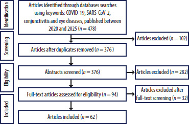
The selection process included screening titles and abstracts (n = 478; after removing 102 duplicates), followed by full-text assessment of 94 articles. After excluding 32 studies meeting the exclusion criteria, 62 publications were included in the analysis (Figure 1).
Figure 1
Preferred Reporting Items for Systematic Reviews and Meta-Analyses (PRISMA) flow diagram illustrating the study selection process
n – number of publications; COVID-19 – coronavirus disease 2019; SARS-CoV-2 – severe acute respiratory syndrome coronavirus 2
From each study, data on authors, country, study type, sample size, observed ocular symptoms, and main findings were extracted. The data were summarized in a synthetic table (Table I).
The data extraction and analysis process was conducted jointly by all authors, with mutual verification at each stage to minimize the risk of systematic error. The analyzed studies involved patients with SARS-CoV-2 infection confirmed using molecular methods (reverse transcription polymerase chain reaction or equivalent diagnostic tests), in whom the presence of ocular symptoms was assessed.
Methodological quality was assessed narratively, considering sample size, presence of a control group, clarity of diagnostic criteria, and the way results were reported. Risk of bias was analyzed in the context of the retrospective nature of some studies, small sample sizes, population heterogeneity, and potential selective reporting. The most common sources of potential systematic error were small sample sizes, the retro- spective design of some studies, and lack of control groups. Additionally, some studies could be affected by selective outcome reporting.
The included studies varied in diagnostic methods (clinical vs. imaging studies), populations (hospitalized vs. outpatient), and definitions of ocular symptoms, limiting the possibility of direct comparison of results. No formal assessment of publication bias was performed due to the absence of a meta-analysis; however, there is a risk of overrepresentation of cases with more severe disease or prominent ocular symptoms.
Due to heterogeneity in methods, populations, and reported outcomes, a qualitative (narrative) analysis was performed without a quantitative meta-analysis. The systematic review method with narrative elements allowed for a synthetic presentation of current knowledge on ophthalmic aspects of COVID-19, despite limitations arising from study heterogeneity and potential publication bias.
The review was not registered in the International Prospective Register of Systematic Reviews (PROSPERO) database due to its narrative nature and limited time frame.
RESULTS
Analysis of 62 publications revealed a wide spectrum of ophthalmic changes in patients with COVID-19, mainly involving ocular surface symptoms and, less frequently, retinal and neuro-ophthalmic alterations.
Table I
Characteristics of studies on ophthalmic manifestations of coronavirus disease 2019 (COVID-19)
| Authors, year | Study design | Population | Ocular findings | Ocular location | Main findings |
|---|---|---|---|---|---|
| Wu P et al., 2020 [2] | Cross-sectional | n = 38 | Conjunctival congestion, epiphora, chemosis | Anterior | Ocular symptoms in 31.6% of cases; more frequent in severe COVID-19 |
| Boz AAE et al., 2021 [3] | Cross-sectional | n = 40 | Redness, tearing, irritation | Anterior | Ocular manifestations in 20% of COVID-19 cases |
| Cavalleri M et al., 2020 [4] | Cross-sectional | n = 43 | Conjunctivitis, dry eye, blurred vision | Anterior | Ocular findings associated with systemic severity and inflammation |
| Nasiri N et al., 2021 [5] | Systematic review & meta-analysis | n = 2,347 (16 studies) | Conjunctivitis, ocular pain, dry eye | Anterior | Pooled prevalence of ocular manifestations ~11%; conjunctivitis ~12% |
| Tran E et al., 2024 [6] | Systematic review & meta-analysis | n = > 8,000 (45 studies) | Conjunctivitis, tearing, dry eye | Anterior | Confirmed ocular symptoms in ~11% of COVID-19 patients; conjunctivitis most common |
| Lima LCF et al., 2021 [7] | Case report | n = 1 | Conjunctivitis in early phase | Anterior | Conjunctivitis reported as early symptom of COVID-19 |
| Gascon P et al., 2020 [8] | Case report | n = 1 | Cotton wool spots, retinal hemorrhages | Posterior | Retinopathy associated with acute COVID-19 infection |
| Gonzalez-Lopez JJ et al., 2021 [9] | Case report | n = 1 | Symptomatic retinal microangiopathy | Posterior | Retinal vascular changes linked to COVID-19 |
| Chen L et al., 2020 [10] | Cross-sectional | n = 535 | Conjunctival congestion, dry eye, blurred vision | Anterior | Ocular symptoms in 5% of cases; mainly conjunctival |
| Kumar KK et al., 2021 [11] | Cross-sectional | n = 1,167 | Conjunctivitis, redness, epiphora | Anterior | Ocular findings in ~7% of patients; more common in moderate/severe cases |
| Bypareddy R et al., 2021 [12] | Prospective | n = 82 | Fundus changes | Posterior | No significant fundus abnormalities in non-severe COVID-19 |
| Dipu T et al., 2022 [13] | Cross-sectional | n = 84 | Retinal vascular changes, microangiopathy | Posterior | Long-term retinal sequelae observed after severe COVID-19 |
| Mocanu V et al., 2022 [14] | Case-control | n = 36 vs. 72 controls | Conjunctivitis as single finding | Anterior | Conjunctivitis identified as lone manifestation of COVID-19 |
| Shroff D et al., 2022 [15] | Case series | n = 7 | Retinal vein occlusion, arterial occlusion | Posterior | Spectrum of retinal vaso-occlusive disorders following COVID-19 |
| Wan KH et al., 2022 [16] | Cross-sectional | n = 288 | Dry eye, ocular surface disturbance | Anterior | Post-COVID-19 ocular surface abnormalities common in convalescent phase |
| Güemes-Villahoz N et al., 2020 [17] | Cross-sectional | n = 164 | Conjunctivitis | Anterior | Conjunctivitis prevalence ~5.5%; mild and self-limited |
| Ozturker ZK, 2021 [18] | Case report | n = 1 | Conjunctivitis as sole symptom | Anterior | Conjunctivitis presenting as isolated COVID-19 manifestation |
| Khavandi S et al., 2020 [19] | Case report | n = 1 | Conjunctivitis | Anterior | COVID-19 presenting as conjunctivitis, highlighting diagnostic challenge |
| Ma N et al., 2020 [20] | Cross-sectional | n = 216 | Conjunctivitis, redness, tearing | Anterior | Ocular symptoms in pediatric COVID-19 patients (~22%) |
| Marinho PM et al., 2020 [21] | Case series | n = 12 | Cotton wool spots, microhemorrhages | Posterior | Retinal changes consistent with microangiopathy in COVID-19 patients |
| Erogul O et al., 2022 [22] | Observational | n = 41 | Retinal microvascular changes | Posterior | COVID-19 associated with retinal microvascular alterations |
| Casagrande M et al., 2020 [23] | Post-mortem | n = 3 (deceased) | Viral RNA in retina | Posterior | SARS-CoV-2 detected in retinal biopsies of COVID-19 patients |
| Mehta S, Pandey A, 2020 [24] | Case report | n = 1 | Rhino-orbital mucormycosis | Orbit | Severe fungal superinfection associated with COVID-19 |
| Ocansey S et al., 2020 [25] | Cross-sectional | n = 38 | Redness, tearing, burning sensation | Anterior | Suggested possible ocular shedding/ transmission of SARS-CoV-2 |
| Abrishami M et al., 2020 [26] | Cross-sectional | n = 93 | Conjunctivitis, dry eye, redness | Anterior | Ocular symptoms present in 22% of hospitalized COVID-19 patients |
| Mishra SK et al., 2024 [27] | Cross-sectional survey | n = 150 (HCWs) | Dry eye, redness, blurred vision | Anterior | Ocular complaints common after COVID-19 infection among HCWs |
| Marquezan MC et al., 2021 [28] | Case report | n = 1 | Conjunctivitis | Anterior | Conjunctivitis in non-severe COVID-19 case |
| Cabot F et al., 2021 [29] | Cross-sectional | n = 29 | Conjunctival hyperemia, chemosis | Anterior | Documented conjunctival changes in COVID-19 cohort |
| Sanjay S et al., 2021 [30] | Case report | n = 1 | Neuro-ophthalmic sequelae | Posterior | Severe ocular complications after recovery from COVID-19 |
| Hösel K et al., 2022 [31] | Observational | n = 89 | Ocular surface irritation, conjunctivitis | Anterior | Documented ocular manifestations in COVID-19 patients |
| Cheema M et al., 2020 [32] | Case report | n = 1 | Keratoconjunctivitis | Anterior | Keratoconjunctivitis was the initial presenting symptom of COVID-19, preceding respiratory manifestations |
The identified studies included both original research (prospective and cross-sectional), systematic reviews and meta-analyses, as well as case reports. Data indicate that ocular symptoms in COVID-19 occur with a frequency of several to more than ten percent, depending on the population and study methodology. The most commonly reported symptoms were conjunctivitis, redness, tearing, dry eye sensation, and itching, with some cases also presenting visual disturbances, eye pain, and posterior segment complaints (Figure 2) [5, 6, 8, 9].
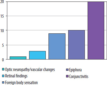
Cross-sectional studies in large populations indicated that changes in the conjunctiva and ocular surface occurred in approximately 11–12% of patients [5, 10, 11]. Less frequently observed manifestations involved the retina – cotton wool spots, retinal hemorrhages, microcirculatory disturbances, and vascular changes [4, 12, 13]. Case reports and small patient series showed that ocular symptoms can be either concomitant or the sole manifestation of COVID-19 [7, 11].
Some studies addressed specific complications, such as rhino-orbital mucormycosis during or after COVID-19 [14], as well as thrombotic retinopathies and vascular complications [15]. Reports also emerged regarding chronic symptoms, including ocular surface dysfunction or long-term retinal microcirculation disturbances following infection [16, 17]. The obtained data suggest that the expression of ACE2 and TMPRSS2 receptors in ocular tissues may provide a biological basis for SARS-CoV-2 tropism. Changes observed in the retina and optic nerve may result from microangiopathy and inflammatory responses secondary to infection.
DISCUSSION
The results highlight significant heterogeneity in the clinical presentation, ranging from mild ocular surface changes to severe complications involving the posterior segment of the eye and the optic nerve.
Ocular surface changes
The most commonly reported ophthalmic manifestation of COVID-19 was conjunctivitis. Meta-analyses reported its prevalence at 11–12% of all patients infected with SARS-CoV-2 [5, 33]. Large clinical cohorts observed typical symptoms such as redness, tearing, burning, itching, foreign body sensation, and eye pain [2, 10, 17]. Notably, in some cases, conjunctivitis was the only or first symptom of infection [18, 19]. This has particular epidemiological significance, as isolated ocular symptoms may indicate the need for diagnostic testing and early identification of infection. A high prevalence of ocular symptoms was reported in the pediatric population, affecting up to one-fifth of children, where conjunctivitis is often the leading sign [20]. In contrast, adult patients, particularly those hospitalized with severe systemic disease, more frequently exhibit posterior segment manifestations and vascular complications. While ambulatory patients typically present with self- limiting ocular surface irritation, hospitalized individuals are at a higher risk for more complex ophthalmic involvement due to both the viral infection and the intensive systemic therapy required [6, 12, 13].
Posterior segment manifestations and vascular complications
Although less frequent, involvement of the posterior segment of the eye has been documented. Clinical observations and imaging studies indicate the presence of retinal microangiopathy, cotton wool spots, petechiae, and other microcirculatory disturbances [12, 21, 22]. Case reports confirmed retinal vascular thrombosis leading to visual impairment [15]. Postmortem studies demonstrated the presence of SARS-CoV-2 genetic material in retinal tissues [23], providing evidence for the potential direct tropism of the virus for ocular structures.
Neuro-ophthalmic and inflammatory complications
In addition to retinal changes, neuro-ophthalmic complications have been reported. These included optic neuro- pathies and neuroinflammatory responses secondary to infection [34, 35]. Experimental data using human retinal organoids demonstrated the virus’s ability to infect and replicate in photoreceptor and ganglion cells, providing further evidence for a direct mechanism of neuro-ocular damage [36].
Severe opportunistic infections
Reports also describe severe secondary opportunistic infections in the orbital region in patients after COVID-19. Rhino-orbital mucormycosis was particularly dangerous, especially in immunosuppressed patients or those treated with steroids [24]. Although these cases are relatively rare, they carry high mortality and highlight the need for heightened clinical vigilance.
Biological mechanisms
The basis for understanding the pathogenesis of ocular symptoms is the expression of ACE2 and TMPRSS2 receptors in ocular tissues – conjunctiva, cornea, and retina [1]. These receptors play a key role in viral entry into cells. Other molecules, such as CD147 (BSG), may also facilitate viral penetration into ocular structures. This supports not only the possibility of direct infection but also the role of the eye as a potential reservoir and site of SARS-CoV-2 transmission [25, 37].
Epidemiological variability and study limitations
The reviewed studies show substantial variation in the prevalence of ocular symptoms, ranging from below 5% to as high as 20% of cases [10, 20, 26]. This discrepancy likely results from methodological differences: some studies relied solely on self-reported symptoms, while others included detailed ophthalmic examinations with a slit-lamp. Limitations of many analyses also included small sample sizes, lack of control groups, and retrospective study design.
Clinical significance
The data indicate that ocular symptoms may be clinically relevant for diagnosing and monitoring patients with COVID-19. On one hand, conjunctivitis and other ocular surface symptoms may be the first or sole manifestation of infection, serving as an early warning sign. On the other hand, observations of retinal and neuro-ophthalmic changes confirm that SARS-CoV-2 can induce serious vascular and inflammatory complications, potentially affecting patient prognosis.
Directions for further research and practical implications
Available systematic and review analyses highlight the need to deepen understanding of mechanisms underlying ocular damage in COVID-19 [38, 39]. Existing data are fragmentary and require standardization in both diagnostic methods and symptom reporting [38]. The interaction of SARS-CoV-2 with ACE2 and CD147 receptors in ocular tissues is emphasized as a foundation for further experimental studies on molecular mechanisms of damage [39]. Other reports highlight the ocular surface as a potential site for both clinical manifestations and viral transmission [40–42]. The conjunctiva and tear film may serve as reservoirs for SARS-CoV-2, with epidemiological significance, particularly for protecting healthcare personnel. Literature also emphasizes that, despite relatively low frequency, ocular symptoms of COVID-19 are practically important for patient care and hospital infection prevention [43].
Population studies indicate that ocular complications may also occur in later stages of the disease, including chronic dry eye, tear film disturbances, and visual impairments, suggesting persistent neuroinflammatory effects or microcirculatory dysfunction [27]. Moreover, “ocular long-COVID” manifestations, such as persistent dry eye and microvascular retinal changes, underscore the need for long-term ophthalmologic follow-up in recovered patients.
Role of ophthalmologists in the pandemic and practical challenges
Ophthalmologists play a key role as first-contact specialists for patients whose ocular symptoms may precede general COVID-19 symptoms [44, 45]. During the early months of the pandemic, special attention was given to diagnostic caution and the use of appropriate personal protective equipment [46, 47]. Even mild conjunctivitis should be treated as potentially infectious, which is crucial for workflow organization in ophthalmology clinics [48].
Wide spectrum of symptoms and clinical consequences
Review and narrative studies confirm that the spectrum of COVID-19 ophthalmic complications is broad, including ocular surface changes, severe vascular and neuro- ophthalmic complications, and opportunistic infections [49–54]. Beyond direct viral damage, the management of severe COVID-19 with high-dose corticosteroids significantly increases the risk of secondary ocular complications, including steroid-induced ocular hypertension, glaucoma, and cataract formation [50]. Moreover, cases of isolated conjunctivitis or singular post-COVID-19 complications demonstrate that SARS-CoV-2 may present solely through ocular symptoms [55–62]. The clinical presentation has evolved with the emergence of different SARS-CoV-2 variants. While earlier strains such as Delta were associated with higher systemic severity, the Omicron variant has been linked to more frequent but milder ocular surface symptoms. Furthermore, the role of COVID-19 vaccination is increasingly recognized; although rare, ocular side effects such as uveitis or retinal vascular events have been reported after vaccination, necessitating careful differentiation from infection-related symptoms [58].
Review findings and quality of evidence assessment
Some analyses have noted issues of publication bias and data overinterpretation. For example, Visioli et al. [60] highlighted erroneous citations and reporting bias in ophthalmic COVID-19 studies. Similar warnings were issued in earlier reports, emphasizing the need for critical assessment of evidence and avoiding excessive generalizations [61, 62].
In summary, although the literature on ocular symptoms of COVID-19 is extensive, it exhibits significant methodological variability. Further high-quality research is needed to better define the pathogenesis, epidemiology, and clinical significance of ophthalmic manifestations in COVID-19.
Most analyses were retrospective; therefore, prospective studies with standardized diagnostic criteria and assessment methods for ocular changes are necessary.
Limitation of the study
This review has several inherent limitations. First, the included studies exhibit substantial heterogeneity in study design, population characteristics, diagnostic methods, and outcome reporting, which limits the ability to perform quantitative synthesis or meta-analysis. Many original studies were retrospective, with small sample sizes, increasing the risk of selection bias and reducing the generalizability of findings. Second, publication bias may have influenced the available evidence, as studies reporting significant or unusual ocular manifestations are more likely to be published. Third, data on long-term ocular outcomes and rare complications remain limited, restricting conclusions regarding chronic or delayed effects of COVID-19 on the eye. Another limitation is the search strategy; by focusing on broad keywords such as “eye diseases” and “conjunctivitis,” some reports concerning very rare or specific ocular complications might have been underrepresented in the initial search results. Finally, the review was not registered in PROSPERO, which may affect transparency, although the methodology strictly followed PRISMA guidelines. Despite these constraints, this systematic review enhances understanding of ocular involvement in COVID-19 and highlights directions for high-quality prospective studies.
CONCLUSIONS
Ocular manifestations of COVID-19, although less frequent than respiratory symptoms, represent an important component of the clinical presentation. They most commonly involve the ocular surface but may also include retinal and neuro-ophthalmic changes. Observed disturbances may result from ACE2 receptor expression in ocular tissues and microangiopathic changes induced by infection. Further prospective studies are necessary to evaluate the long-term ophthalmic consequences of COVID-19. Understanding the ocular aspects of COVID-19 is significant not only diagnostically but also epidemiologically, enabling early identification of infection and reduction of viral transmission.