CASE STUDY
A 40-year-old Caucasian male patient with no prior ocular or medical history presented to the ophthalmic hospital due to a foreign body sensation in the right eye that had persisted for several weeks. The patient did not report any deterioration of vision or any other symptoms; however, the affected eye was amblyopic. He denied any trauma to the eye or contact lens usage.
On examination, the visual acuity in the right eye (visus oculus dexter, VOD) was 0.16 on the Snellen chart, with no improvement upon correction. The patient was unable to read the Snellen near vision chart with this eye. Intraocular pressure (IOP), measured by Goldmann applanation tonometry, was 12 mmHg. Slit-lamp examination revealed central corneal edema and a foreign body in the conjunctival sac, which was subsequently removed. Visualization of the deeper anterior segment structures was limited because of the corneal edema. Central corneal erosion was also found in slit-lamp examination. The left eye was unremarkable. The patient was administered ofloxacin ointment to the right eye 4 times daily and at bedtime and advised to return for a follow-up visit in 5 days or earlier if further visual deterioration occurred.
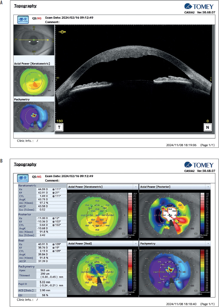
Five days later, the patient returned for a follow-up, reporting an improvement in symptoms, including a reduced foreign body sensation. However, the cornea remained hazy and thickened, with only a narrow, clear zone adjacent to the limbus; no conjunctival hyperemia was found. Pachymetry of the right cornea revealed a central corneal thickness (CCT) of 1200 μm (Figures 1A and 1B). In contrast, the left eye demon- strated normal distance and near (Snellen) visual acuity, with an IOP of 12 mmHg and CCT of 545 µm. Following the adjustment in diagnosis, the patient’s treatment regimen was revised to include topical hyperosmotic saline solution (four times daily), dexamethasone (twice daily) and systemic acetazolamide (250 mg twice daily), and potassium chloride (391 mg once daily) as symptomatic treatment for post- traumatic corneal edema.
Figure 1
A) Anterior segment optical coherence tomography (AS-OCT) scan revealing central corneal edema with marked corneal thickening. B) AS-OCT corneal analysis maps of the right cornea showing marked corneal thickening, with a paracentral corneal thickness of 1189 µm
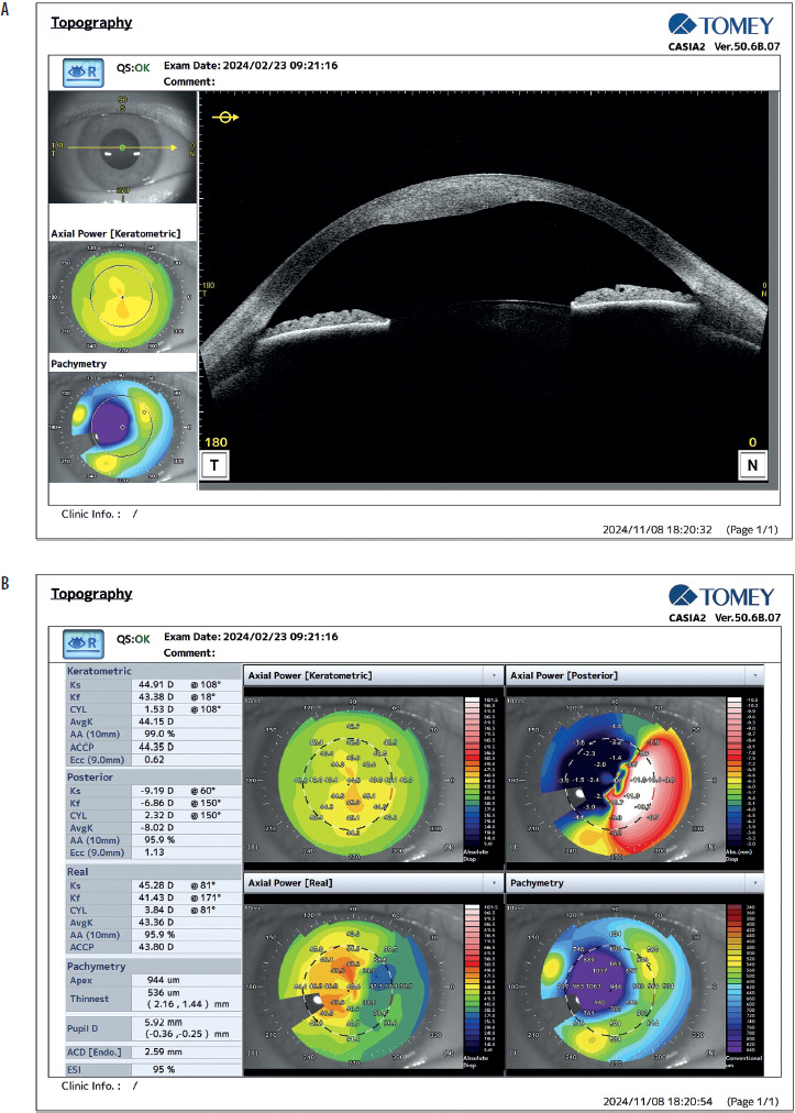
At the follow-up visit (the third visit), VOD improved to 0.4 on the Snellen chart. Slit-lamp examination revealed persistent central corneal edema but with clinical improvement (Figure 2). The presence of corneal edema in the right eye was further confirmed using keratometry (Figures 3A and 3B). However, the persistence of corneal edema, together with signs of inflammation, suggests that the empirical treatment was not optimal.
Figure 2
Slit-lamp examination showing persistent, but reduced, central corneal edema at the follow-up visit
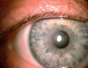
Figure 3
A) Anterior segment optical coherence tomography scan showing a reduction in central corneal edema. B) Corneal topography scan showing a slight reduction in central corneal thickness
The persistence of corneal edema and inflammation despite appropriate symptomatic therapy prompted consideration of alternative diagnoses causing secondary corneal edema, including iridocorneal endothelial syndrome (ICE) and unilateral corneal dystrophy. Unilateral corneal edema without elevated IOP, together with a normal contralateral eye, suggests endothelial decompensation associated with ICE in the affected eye. This condition is known for its asymmetric presentation [1]. To test this hypothesis, advanced diagnostic evaluations were planned, including confocal microscopy (CONFOSCAN, in vivo confocal microscopy), gonio- scopy, and anterior segment optical coherence tomography (AS-OCT). Gonioscopy indicated an open angle with visible structures extending to the ciliary body, without any pathology within the drainage angle. Confocal microscopy suggested the presence of atypical endothelial cell morphology consistent with ICE in the right eye; the median endothelial cell count was within normal limits (2890 cell/mm2). AS-OCT revealed a corneal thickness measurement of 1063 μm.
Following revision of the working diagnosis, the patient’s treatment regimen was revised to include topical hyperosmotic saline solution (four times daily), dexamethasone (six times daily) and systemic acetazolamide (250 mg twice daily), and potassium chloride (391 mg once daily). The aim of this combination was to reduce corneal edema.
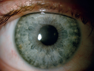
During the subsequent visit, the patient’s condition showed overall signs of deterioration. VOD had declined to 0.32 sine correctione (sc), while near visual acuity had decreased to 1.5 sc. The central corneal edema persisted, although its extent was reduced compared with the previous visit (Figure 4). Additionally, fine stellate keratic precipitates were visible on the endothelium surrounding the edema in an approximately 1 mm ring.
These findings highlight the progressive nature of the patient’s condition, the persistence of corneal edema, and the appearance of endothelial deposits.
The results from CONFOSCAN, AS-OCT, and gonioscopy provided crucial diagnostic information, and further supported consideration of ICE syndrome. However, the presence of fine non-granulomatous deposits on the endothelium and central corneal edema could also indicate herpes simplex keratitis (HSK), as well as other infections such as toxoplasmosis and other syndromes, including Fuchs heterochromic iridocyclitis [2]. HSK can present with similar clinical features, including dendritic lesions and endothelial inflammation, which may mimic ICE [3]. In order to control these conditions, the following medications were administered: topical dexamethasone ophthalmic drops (six times daily) and systemically: acyclovir (dose 5 × 800 mg orally), methylprednisolone (24 mg once daily orally) and omeprazole (20 mg once daily). The aim of this therapeutic approach was to address both the potential viral etiology and the inflammatory component of the patient’s condition.
At the one-week follow-up visit, the patient exhibited notable improvement. With correction of the right eye at –2.25 diopters, VOD improved to 0.5 on the Snellen chart, and near visual acuity (Snellen near chart) was 0.5. IOP slightly increased to 14 mmHg. Although central corneal edema persisted, its severity was substantially reduced compared with the previous week, with AS-OCT indicating a thickness of 615 μm (Figure 5). Given the patient’s improving condition, a follow-up appointment was scheduled for two weeks’ time to monitor further progress.
Figure 5
Anterior segment optical coherence tomography scan showing mild central corneal edema; central corneal thickness of 615 µm was found
This clinical progression suggested a favorable response to treatment. Improvements in both pachymetry measurements and visual acuity were accompanied by a progressive reduction in corneal edema.
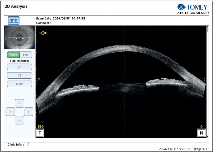
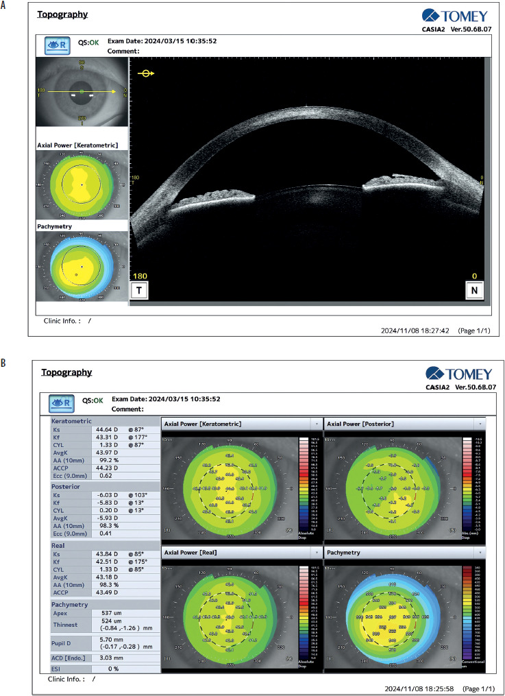
Upon the patient’s return for a follow-up visit two weeks later, further improvement was observed. To achieve equivalent visual acuity in both distance and near vision (VOD = 0.5, Snellen = 0.5) in the right eye, a reduced correction of –1.75 diopters was necessary. Slit lamp examination revealed a significant reduction in corneal edema in the central zone, with diminishing haziness. Additionally, AS-OCT demonstrated a decrease in corneal thickness to 537 μm (Figures 6A and 6B).
DISCUSSION
Final diagnosis
The patient’s condition improved significantly following the administration of oral acyclovir and extensive topical dexamethasone. This therapeutic response, together with the clinical findings, strongly supported a diagnosis of HSK. HSK is a viral infection of the cornea caused by the herpes simplex virus (HSV), characterized by recurrent episodes of corneal inflammation and ulceration. In the present case, the improvement in corneal edema and the reduction of visual symptoms following antiviral and immunosuppressive therapy were consistent with this diagnosis.
HSK can present with central corneal edema, dendritic lesions, and endothelial inflammation, which can mimic other corneal pathologies such as ICE. The presence of fine precipitates on the endothelium and the persistence of central corneal edema were initially suggestive of the inflammatory process; however, the significant clinical improvement observed after antiviral therapy, coupled with the decrease in corneal thickness and the normalization of IOP, is more consistent with HSK. Additionally, the absence of elevated IOP and the rapid resolution of symptoms following acyclovir administration favored HSK over ICE syndrome. In contrast to HSK, ICE syndrome typically follows a more chronic course and is not expected to demonstrate such a rapid therapeutic response.
However, the complex presentation, including persistent corneal edema and fine non-granulomatous deposits on the endothelium, raises the possibility of a concomitant diagnosis of ICE syndrome, potentially associated with HSV infection. Studies such as Li et al. [4] have proposed that ICE syndrome’s etiological mechanism may involve HSV infection and the integration of viral genes into the human genome. Detection of HSV DNA in the aqueous humor of ICE syndrome patients by Alvarado et al. [5] and Groh et al. [6] further supports this viral etiology. However, given the strong clinical improvement on topical and oral treatment, aqueous humor sampling was considered inappropriate in the present case. The potential diagnostic benefit of an invasive procedure was considered to be outweighed by its associated risks and unlikely to alter clinical management.
In conclusion, the patient’s positive response to acyclovir therapy, characterized by reduced corneal edema and improved visual acuity, strongly supported the diagnosis of HSK. Nevertheless, the possibility of concurrent ICE syndrome with HSV cannot be excluded. This case underlines the importance of considering viral etiologies in patients presenting with corneal edema and highlights the need for targeted antiviral treatment in managing HSK.
Differential diagnosis
Severe corneal edema was suspected to be secondary to trauma or another underlying cause. Differential diagnoses considered were: a) unilateral corneal dystrophies such as Fuchs’ endothelial dystrophy and posterior polymorphous dystrophy; b) iridocorneal syndromes such as Chandler syndrome (a subtype of Cogan-Reese syndrome); c) uveitis-glaucoma-hyphema (UGH) syndrome; d) infectious keratitis, including bacterial and viral etiologies; and e) corneal decompensation secondary to infection, trauma, or surgery.
Fuchs’ endothelial dystrophy shows similarities to the present case concerning corneal endothelial changes and edema. However, Fuchs’ dystrophy typically manifests bilaterally and mainly affects elderly individuals. Diagnostic modalities such as confocal microscopy and specular microscopy reveal guttae and endothelial cell loss characteristic of Fuchs’ dystrophy [7]. Similarly, posterior polymorphous corneal dystrophy predominantly presents bilaterally. Suspected cases can be confirmed through genetic testing and corneal imaging, which exhibit characteristic endothelial changes [8].
Chandler syndrome (a subtype of the iris nevus Cogan-Reese syndrome) warrants consideration due to its association with corneal edema and abnormal corneal endothelium. However, Chandler syndrome is distinguishable by characteristic iris changes, including iris nodules and peripheral anterior synechiae [9]. Another differential diagnosis is UGH syndrome, which may manifest with corneal edema, elevated IOP, and inflammation secondary to intraocular lens complications. This condition can be easily discerned, as it is associated with intraocular lens placement and can be identified through anterior segment imaging [10].
Finally, unilateral corneal dystrophy was considered because of its similarity to the clinical presentation of the patient. However, corneal dystrophies are typically bilateral hereditary disorders, and truly unilateral cases are rare. In the absence of supporting clinical or family history findings, this diagnosis was considered less likely and remained a diagnosis of exclusion. The patient had no history of ocular surgery.