INTRODUCTION
Laser peripheral iridotomy (LPI) is an established therapeutic method used for the prevention of acute attacks of primary angle-closure glaucoma (PACG). He et al. [1], based on an analysis of nearly 12,000 individuals, demonstrated that LPI significantly reduces the risk of acute glaucoma attacks in eyes with a narrow filtration angle (FA). Gupta et al. [2] reviewed the standard use of 2% pilocarpine before the onset of the “LPI era”. The authors emphasized that, when an acute glaucoma attack is suspected in eyes in which LPI has not been performed, pilocarpine remains an important agent in both treatment and prophylaxis.
The approximate width of the FA can already be determined during an optometric examination using the van Herick method. In this technique, peripheral anterior chamber depth is graded by comparing the thickness of the peripheral cornea with the diameter of the space between the posterior corneal surface and the anterior iris surface [1–3]. The width and morphology of the FA are routinely assessed by gonioscopy [4]. More recently, anterior-segment optical coherence tomography (AS-OCT) has emerged as a non-contact imaging modality for FA assessment. AS-OCT provides several advantages. Notably, it does not require an ophthalmologist to be physically present. The use of AS-OCT in the diagnosis of narrow FAs considerably facilitates the diagnostic process while also allowing remote interpretation of the results obtained.
Until recently, gonioscopy was regarded as the gold standard for FA assessment. However, in view of advances in imaging techniques, the term “standard” is often time-dependent and therefore limited. Just over 20 years ago, extracapsular surgery was the standard technique for cataract surgery, and the lens had to be completely opacified. Today, this technique is considered outdated and is no longer regarded as the standard.
With the introduction of AS-OCT for FA assessment, the question arises as to whether gonioscopy is still considered the most important and optimal examination method. In 2007, Noal et al. [5] demonstrated the effectiveness of AS-OCT in assessing FA closure, with an accuracy of 98%. Imran et al. [6], assessing the FA in cases of FA closure or suspected FA closure, demonstrated greater specificity, at 66.7%, using AS-OCT compared with 33.3% when FA was assessed by gonioscopy. In contrast, Yaisiri et al. [7], comparing gonioscopy and AS-OCT in the diagnosis of FA closure, found virtually no differences in detection sensitivity. The authors also highlighted the benefits of non-contact AS-OCT, which allows the FA to be assessed and visualized both qualitatively and quantitatively.
Important diagnostic features that may indicate an abnormal FA width include ocular pain and/or frontoparietal headache. Ocular pain is one of the non-specific symptoms accompanying an acute glaucoma attack. Other symptoms include headache, blurred vision, seeing halos around light sources, and vomiting. The clinical picture is complemented by pathological changes visible in the anterior segment of the eye, including corneal epithelial oedema, practically complete obliteration of the anterior chamber, and pupillary dilation. The clinical picture of an acute FA-closure glaucoma attack is further characterized by the absence of a pupillary response to light. Ocular symptoms occur when intraocular pressure (IOP) reaches pathologically high values of 50–70 mmHg. It is worth noting that, in this situation, performing LPI does not produce the expected result, i.e., a sustained reduction in the IOP.
Therefore, when an acute glaucoma attack is diagnosed, topical and systemic pharmacological treatment is initiated first (mannitol, acetazolamide, pilocarpine, and drugs that reduce aqueous humor production and increase its outflow) to lower the IOP and thereby improve the conditions for possible LPI or surgical treatment.
As a standard approach, when an acute glaucoma attack is diagnosed in one eye, prophylactic LPI should be considered for the fellow eye.
A narrow FA is not always associated with a shallow anterior chamber. In clinical practice, the opposite situation is also observed, i.e., despite the presence of a deep central ante- rior chamber, there is marked peripheral FA narrowing. This anatomical configuration may be overlooked during routine slit-lamp examination. Typical examples include plateau iris syndrome, eyes with a short anteroposterior axial length, and eyes in which the lens is positioned more anteriorly [8–10]. In these conditions, the van Herick method may yield false-negative results, suggesting a normal FA width, because this method does not take into account the configuration of the entire FA.
To accurately assess FA width and guide further therapeutic management, more detailed diagnostic evaluation is required, including gonioscopy according to the Spaeth classification, which takes into account the location, shape, and relationships of the FA structures [11], and/or AS-OCT, which enables assessment of the iris and FA configuration [12–14]. Diagnosis of a narrow FA in such cases is clinically important, both in the context of prevention of PACG and when making decisions regarding LPI.
According to the recommendations of the European Glaucoma Society, periodic assessment of the FA should be performed in individuals over 40 years of age regardless of the subjective assessment of anterior chamber depth in the central and peripheral parts [14]. It is estimated that marked FA narrowing may be present in approximately 30% of eyes despite the presence of a deep anterior chamber [12, 15]. Furthermore, ocular pain or headache are not always present as prodromal symptoms preceding an acute glaucoma attack.
Example 1. A woman, 44 years; low-grade hyperopia; status post acute glaucoma attack in the right eye
The patient presented to the Silesian Eye Treatment Center in Żory for surgical management of complications resulting from a previous acute glaucoma attack, including removal of a complicated cataract and iridoplasty required because of irreversible damage to the pupillary sphincter.
Before the acute glaucoma attack, the patient had reported neither ocular pain nor frontal headaches. On the day of the acute glaucoma attack, following presentation to the ophthalmic emergency department, LPI was immediately performed in the fellow eye because of the presence of a narrow FA.
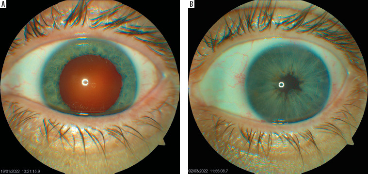
In addition to cataract extraction and implantation of an asymmetric bifocal intraocular lens, the patient underwent surgical reduction of pupil diameter (Figure 1A, 1B).
Figure 1
Color photographs of the anterior segment of the right eye: A) following an acute glaucoma attack, demonstrating an irreversibly dilated pupil; B) after surgical pupillary constriction via a cerclage suture placed at the pupillary margin – pseudophakic eye
Ocular pain or headache reported by patients, particularly frontal headache, should prompt physicians or optometrists to investigate its cause more thoroughly, even when the IOP and visual acuity are within normal limits (author’s note).
Example 2. A woman, 42 years; status post acute glaucoma attack in the right eye; low-grade hyperopia in both eyes
Similar to the patient described in Example 1, this patient presented to the Silesian Eye Treatment Centre in Żory to minimize the sequelae of a previous acute glaucoma attack. For three years, the patient had undergone regular ophthalmic examinations every six months because of non-specific ocular pain and frontal headaches. During these visits, visual parameters remained normal and the IOP did not exceed 15–16 mmHg. Consequently, no additional diagnostic evaluation was undertaken to determine the cause of the reported symptoms.
During hospitalization, LPI was performed in the fellow eye because of a narrow FA (author’s note).
Both cases shared several common features, including normal visual acuity, normal IOP, and the presence of low-grade hyperopia.
AIM OF THE STUDY
The aim of this study was to evaluate the post-procedural outcomes of LPI in patients with narrow FAs and in patients reporting chronic ocular pain and/or frontotemporal headaches, regardless of FA width. In addition, associations between the outcomes of the procedure and selected other factors, including the indication for LPI, sex, ocular anatomy, refractive error, and patient age, were assessed.
MATERIAL AND METHODS
Between 2018 and 2024, more than 700 LPI procedures were performed at a private medical facility, the Silesian Eye Treatment Center in Żory, Poland. A subset of these procedures was included in the present analysis. All procedures were performed by three ophthalmologists.
The standard diagnostic procedure used at the Center included an initial biomicroscopic assessment of the anterior segment of the eye by optometrists while preparing patients for ophthalmic consultation or during optometric examinations performed for refractive error correction. The assessment focused on anterior chamber configuration and FA evaluation using the 4-grade van Herick method. A shallow anterior chamber and a narrow FA graded 0–2 according to the above classification constituted indications for further diagnostic evaluation. When a narrow FA was suspected, patients were asked about ocular pain and/or headache, and other accompanying symptoms.
If ocular pain or frontoparietal headache was reported as the primary reason for the ophthalmic and/or optometric visit, in addition to the standard examination of the anterior segment of the eye, diagnostic assessment of FA width was recommended.
In patients reporting headaches, neurological-type visual field testing was routinely ordered.
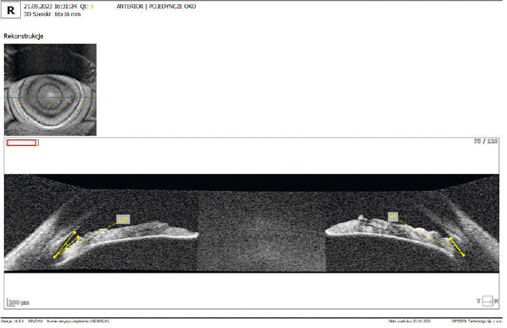
Gonioscopy, an established method of assessing the FA, requires the involvement of an ophthalmologist. Therefore, AS-OCT was performed in nearly all cases to evaluate FA width and morphology. The FA was visualized at two locations by analyzing horizontal scans obtained at the 3 and 9 o’clock positions (Figure 2). In all cases, AS-OCT examinations were performed using a Revo NX 130 device (Optopol Technology). Identification of a narrow FA on AS-OCT constituted the basis for qualification for LPI.
Figure 2
Anterior segment optical coherence tomography scan obtained at two meridians: 3 and 9 o’clock (right eye). Marked filtration angle narrowing is visible at both assessed locations
As a routine component of the optometric and ophthalmic examination, the medical history focused on potential ophthalmic causes of the reported pain, including glaucoma. Patients were also asked about previous neurological, otolaryngological, and dental evaluations undertaken to determine the source of their symptoms. If no ophthalmic cause was identified, patients were referred to the appropriate specialists.
IOP was measured using two methods. First, tonometry was performed using an iCare device or a non-contact air-puff method, with correction for corneal thickness. This was followed by applanation tonometry.
Eligibility criteria for LPI:
patients with no history of previous ophthalmic procedures, either surgical or laser;
identification of a narrow FA constituting an indication for PACG prophylaxis;
presence of ocular pain and/or frontal headache despite a moderately wide FA, with LPI indicated to reduce or eliminate symptoms (in such cases, exclusion of extraocular causes was crucial);
chronic ocular pain and/or frontotemporal headache despite a wide FA (> 30°), after ophthalmic, neurological, otolaryngological, and dental causes had been excluded; this group frequently included patients with large pupils measuring 5.5–6.0 mm in diameter.
All patients were informed about the rationale for the proposed laser procedure, its associated benefits, and possible post-procedural complications. Each patient provided written informed consent to undergo the laser procedure and to the use and processing of their examination results for scientific purposes.
Procedure steps:
IOP measurement before the procedure;
instillation of 2% pilocarpine hydrochloride into the conjunctival sac 2–3 times at 5-minute intervals;
a single instillation of 0.5% proxymetacaine hydrochloride immediately before the procedure;
placement of a gel-filled focusing contact lens for LPI on the anaesthetized eye;
performance of LPI using a neodymium:YAG laser (Nd:YAG, Ellex®) in the superior peripheral portion of the iris, at the site of lowest tissue density;
instillation of an antiseptic preparation;
IOP measurement 10–20 minutes after the procedure;
all patients were instructed to use antiseptic eye drops three times daily for 7 days after the procedure.
A follow-up visit was routinely scheduled 2–3 weeks after the procedure. Patients were advised to return for an unscheduled visit if any symptoms developed in the treated eye.
STATISTICAL ANALYSIS
Statistical significance was set at p = 0.05. Non-parametric tests used for statistical analysis included the Friedman ANOVA and the Mann-Whitney U tests. Spearman’s rank correlation coefficient was used to assess associations between variables. Statistical analyses were performed using R software (version 3.6.0), PSPP, and Microsoft Office 2019.
RESULTS
Information
When interpreting the results obtained, particular attention should be paid to the fact that the analysis employed a classification system in which a single patient could be assigned to more than one category of characteristics. Therefore, the analyzed groups are not mutually exclusive. Accord- ingly, the percentages presented represent the frequency of occurrence of individual characteristics within the study population and should not be interpreted as components that sum to 100%. Consequently, the sum of the percentages across individual categories may exceed 100%, reflecting the possibility that multiple characteristics may occur simultaneously in the same patient. This does not indicate an error in the analysis.
Patient-reported symptoms
During the optometric and medical interviews, the most frequently reported symptoms in the study group were headaches (64.4%), bothersome ocular pain (36.4%), and other symptoms, including photophobia, transient visual loss, and nausea (15.0% of participants). Multiple symptoms frequently co-occurred in the same patient. Only 27.3% of patients reported no symptoms.
Other typically ophthalmic reasons prompting further diagnostic evaluation included deterioration of distance and near visual acuity, blurred or hazy vision, diplopia, distorted (wavy) vision, and the need for preventive follow-up examinations.
A very commonly reported complaint was ocular discomfort, encompassing a painful pressing sensation in the eyes, retrobulbar pressure, transient vision loss, photophobia, and severe headaches accompanied by nausea. The symptoms described above were reported by 77.1% of the study group.
Age of patients
The youngest patient was a 10-year-old girl, whereas the oldest was an 89-year-old woman who reported episodes of transient vision loss and ocular pain, occurring mainly in the evening. The characteristics of the study group are presented in Table I.
Table I
Characteristics of the study group, including age, sex, refractive error, reasons for visit, head and ocular symptoms, anterior segment optical coherence tomography (AS-OCT), presence of cataract, and follow-up period
LPI and acute glaucoma attacks
Between 2018 and 2024, no cases of acute glaucoma attack were recorded in eyes that had undergone LPI.
LPI and ocular pain and/or headache
Complete resolution of ocular pain and/or headache was reported by 47.8% of patients. Most of the group consisted of patients with a narrow FA. The study group also included patients with moderately wide and wide FAs (> 20°), many of whom had pupil diameters exceeding 5.5 mm.
An important eligibility criterion for LPI, consistently emphasized in patients with moderately wide and wide FAs, was the absence of pathology on neurological, otolaryngological, and dental examinations, as well as exclusion of other causes of the pain. Symptoms had often persisted for many years, and many patients had previously been treated following a diagnosis of migraine.
A reduction in all symptoms after the procedure, including headaches and ocular pain, nausea, photophobia, and other complaints, was reported by 64.3% of patients. In 15.3% of patients, the symptoms reported before the procedure did not resolve.
Two noteworthy cases involved very young patients, an 18-year-old and the aforementioned 10-year-old girl. Despite repeated ophthalmic examinations and an almost one-month diagnostic stay in a neurology department, the cause of their chronic recurrent headaches could not be established. However, the reported symptoms resolved almost immediately after bilateral LPI had been performed following confirmation of narrow FAs (author’s note).
IOP (mmHg) – mean values obtained before and after LPI
Before the procedure, the mean IOP was 18 mmHg in the right eye (range: 11–24 mmHg) and 17.95 mmHg in the left eye (range: 11–25 mmHg). At the follow-up examination performed 2–3 weeks after the procedure, the mean IOP was 15 mmHg in the right eye (range: 6–22 mmHg) and 15.46 mmHg in the left eye (range: 6–23 mmHg), respectively. Following LPI, the mean IOP decreased by 2.5–3.0 mmHg, depending on the eye.
Sex and LPI
Women constituted the majority of the study group (77.9%), although eligibility for the procedure was determined by FA width and reported pain.
Refractive error and LPI
Patients with hyperopia constituted the majority of the study group (78.2%). As with sex, the decision to perform the procedure was based on the presence of narrow FAs and reported pain.
Narrow FA was incidentally detected during the first visit in 94.1% of patients. Notably, most of these patients had previously undergone ophthalmic examinations, often on multiple occasions. However, only 5.5% of the entire group had been referred for LPI by physicians not affiliated with the center. Diagnostic evaluation for glaucoma was the reason for the visit in only 15.5% of patients in this group.
A series of analyses was conducted to evaluate the relationships among ocular anatomical parameters, FA width, sex, age, and refractive error. The results are presented below.
Analysis 1. Is there a difference in FA width between measurements taken from the nasal and temporal sides?
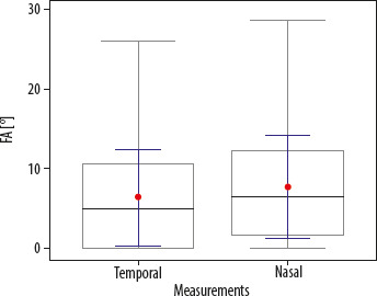
Results: The median FA width was 5.00° for measurements on the temporal side and 6.50° for measurements on the nasal side. The difference was statistically significant (p < 0.05). FA values are illustrated in the box plot (Figure 3).
Figure 3
Graph showing mean filtration angle (FA) values measured temporally and nasally. Statistically significant smaller FA values were observed on the temporal side
Given the percentage distribution of FA width values, the analysis was performed separately for the right and left eyes. The values obtained are presented in Table II.
Table II
Percentage distribution of filtration angle (FA) width, analyzed separately for the right eye and left eye
| Variable | Category | n | % |
|---|---|---|---|
FA – right eye | Closed | 95 | 37.5 |
| 1–5° | 32 | 12.6 | |
| 6–10° | 43 | 17.0 | |
| > 10° | 46 | 18.2 | |
| No information | 37 | 14.6 | |
FA – left eye | Closed | 105 | 41.5 |
| 1–5° | 31 | 12.3 | |
| 6–10° | 33 | 13.0 | |
| > 10 ° | 49 | 19.4 | |
| No information | 35 | 13.8 |
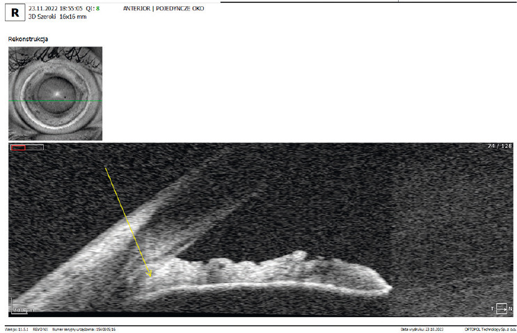
The results indicated complete FA closure in 37.5% of cases in the right eye and 41.5% of cases in the left eye (Figure 4). This finding was generally limited to a single measurement, predominantly on the temporal side. FA values ≤10° were found in 67.1% of cases in the right eye and 66.8% of cases in the left eye.
Figure 4
Anterior segment optical coherence tomography scan illustrating the filtration angle (FA) in right eye. The scan indicates complete FA closure
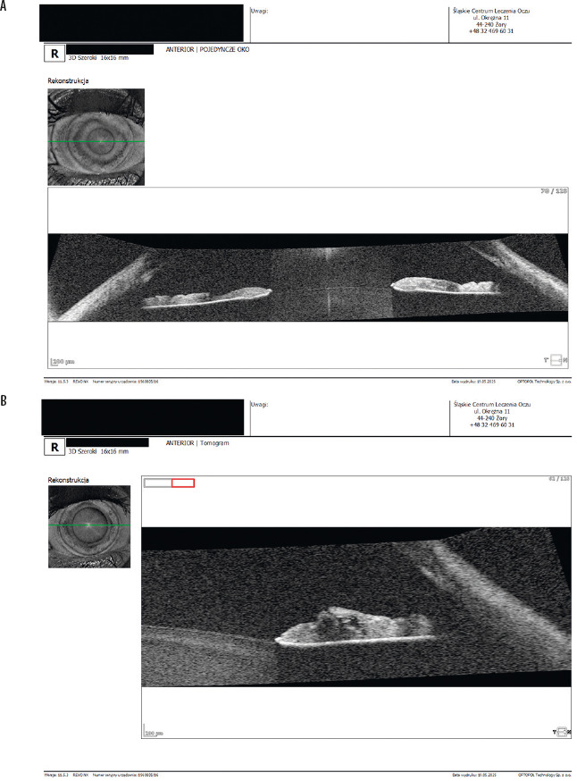
In 14.6% of cases in right eyes and 13.8% of left eyes, FA assessment by AS-OCT was not possible because of an ambiguous image. This was caused by a shadow cast by the sclera onto the FA apex (Figure 5).
Figure 5
Anterior segment optical coherence tomography scan illustrating filtration angle (FA) configuration: A) evaluated at two meridians, B) evaluated at a single meridian. Individual anatomical FA structures could not be visualized because of shadowing cast by the scleral spur
No correlation was demonstrated between pupil diameter and FA width. Also, no association was observed between age and the reduction or complete resolution of pain.
Analysis 2. Are refractive error, its magnitude, pupil diameter, age, and sex significant predictors of FA width?
Results: The analysis revealed no significant associations between FA width and refractive error, its magnitude, pupil diameter, age, or sex.
Analysis 3. Are refractive error, its magnitude, pupil diameter, age, and sex significant predictors of resolution or persistence of symptoms?
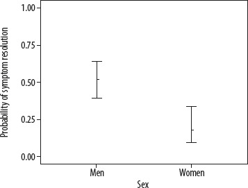
Results: A detailed analysis of the regression coefficients demonstrated a statistically significant effect of sex on symptom resolution. The regression coefficient for sex was negative. Odds ratio (OR) analysis showed that the odds of symptom resolution in men were 79% lower than in women (OR = 0.21).
Table III presents the probabilities of symptom resolution according to sex.
Table III
Probabilities of symptom resolution according to sex
| Sex | p | SE | 95% confidence interval | |
|---|---|---|---|---|
| Lower limit | Upper limit | |||
| Women | 0.52 | 0.06 | 0.40 | 0.63 |
| Men | 0.19 | 0.06 | 0.09 | 0.34 |
Among women, the probability of symptom resolution was p = 0.52, whereas among men it was significantly lower, at p = 0.19. Thus, symptoms resolved significantly more frequently in women. This finding supports the initial hypothesis regarding the effect of sex on symptom resolution (Figure 6).
Pupil diameter vs. FA width, and patient age vs. symptom resolution
No statistically significant associations were found between pupil diameter and FA width, nor between patient age and the reduction or complete resolution of symptoms.
Procedural complications
Intraoperative
Bleeding from the LPI site occurred in 122 eyes. It resolved after increasing pressure on the globe for 20–30 seconds using the contact lens employed during the LPI procedure.
Immediately postoperative
An increase in the IOP above 25 mmHg was noted in 97 eyes. Depending on the IOP values, patients were administered topical IOP-lowering medications. When the IOP exceeded 30 mmHg, oral acetazolamide was administered additionally.
During the subsequent postoperative period
Keratitis occurred in 1 eye, whereas conjunctival irritation was reported in 43 eyes. Patients also reported photophobia, dizziness, transient visual disturbances, moving dark spots in the visual field, headaches, ocular pain, mild self-limiting conjunctival inflammatory reaction, and glare. These complaints resolved within several days to two weeks after the procedure.
DISCUSSION
The results of the present study indicate an association between FA anatomy and the occurrence of ocular pain and headache, as well as their resolution following LPI. As indicated by the statistical analysis, a reduction or complete resolution of pain was observed in 84.7% of patients.
LPI is performed in both adults and children reporting frontal headache or ocular pain. Teixeira et al. [16] performed prophylactic LPI in 60 eyes with a narrow FA and reported resolution of headache in 79.2% of cases, highlighting the need for further studies to investigate the potential association between narrow FAs and headaches. Pinto Ferreira et al. [17] described the case of a 13-year-old patient whose headaches, which had persisted for more than six years, resolved following LPI. Although LPI in children is considered controversial by some physicians, the findings of that study indicate that patient age alone should not be the sole factor precluding the possibility of performing this procedure. Based on an analysis of 20 patients younger than 18 years (38 eyes), Sponsel et al. [18] described atypical FA configurations observed on gonioscopy. These unusual changes mainly included marked stretching of the iris during gonioscopic compression and concentric linear iris pigment deposition anterior to the trabecular meshwork. According to the authors, such anatomical features may play a role in the persistence of headaches and their resolution following LPI, despite normal IOP values and visual acuity. The authors of that study also highlighted an association between excessive accommodation, exposure to bright illumination, and recurrent frontal headaches.
Analysis of the study results points to one important aspect of the diagnostic process that warrants critical consideration: although it was beneficial for the patients studied, it also highlighted shortcomings in their previous diagnostic evaluation. Narrow FAs were detected incidentally in more than 94% of patients despite the fact that most of them had undergone ophthalmic examinations on multiple occasions.
This finding may indicate that FA assessment had not been given sufficient attention during previous diagnostic evaluations, which focused primarily on normal visual acuity and IOP values. When asked whether anyone had previously informed them that their ocular anatomy might predispose them to a marked increase in the IOP, most patients responded negatively. The exceptions were patients referred because of suspected or confirmed glaucoma. However, these individuals accounted for only 15.4% of the study population.
The mean IOP measured 2–3 weeks after LPI was 2.5–3 mmHg lower than pre-procedural values, which may indicate an association between narrow FAs and the IOP.
A positive aspect of the study is that optometrists, as a professional group often serving as the first point of contact for patients, evaluated FA width as part of their routine examinations, thereby prompting further assessment. However, this level of diagnostic evaluation requires an appropriate level of training.
Identification of narrow FAs was a factor prompting a more extensive optometric and ophthalmic history. Ocular pain was reported by 36.4% of patients, whereas 64.4% of the study participants reported frontotemporal headache.
CONCLUSIONS
Performing LPI, in addition to its potential protective effect against acute glaucoma attacks, was associated with a reduction or complete resolution of ocular pain and/or headache in a substantial proportion of the patients studied, including those without unequivocally narrow FAs. The procedure was performed more frequently in women and in individuals with hyperopia, despite FA width being the primary eligibility criterion. In patients with chronic ocular pain and/or frontotemporal headache, after extraocular causes of the symptoms had been excluded, LPI was associated with a reduction or complete resolution of symptoms also in patients with normal FA width, including pediatric patients.
A thorough assessment of the anterior segment of the eye, including an initial assessment of FA width using the van Herick method supplemented by AS-OCT, may facilitate the dete- ction of narrow FAs and reduce the risk that they will be overlooked.