INTRODUCTION
With approximately 10 million cases globally and 1.4 million deaths, tuberculosis (TB) continues to be a serious health issue that warrants urgent attention from health-care systems. Most TB cases occurred in underdeveloped nations. Indonesia ranks second in the world in terms of TB burden, with a projected 969,000 cases overall and a prevalence rate of 354 cases per 100,000 individuals [1]. Ethambutol continues to be used extensively as the first-line treatment for TB (often in combination with other medications). Although this medication is generally well-tolerated by patients, aftereffects have been recorded since its introduction for TB treatment, the most concerning of which is ethambutol toxic optic neuropathy (ETON). One study reported a frequency of 2.25% (22.5 incidences of toxic optic neuropathy per 1000 patients), whereas other studies have reported a frequency of up to 35% [2–4].
Clinically, ETON is characterised by bilateral and asymmetric dyschromatopsia, painless loss of central vision, and decreased contrast sensitivity. Although bitemporal hemianopia has also been reported, a central or cecocentral scotoma is the more common visual field impairment [5]. Generally, symptoms start appearing 4–12 months after ethambutol initiation [6]. Furthermore, despite adequate observation, ETON may develop; therefore, the ocular damage may be irreversible and result in blindness [7]. This case series presents four patients who developed optic neuropathy during ethambutol treatment. These cases highlight several key issues, including an atypical case of bitemporal hemianopia and variability in visual outcomes despite uniform management strategies. Notably, all patients received ethambutol for longer than the duration recommended by the national TB treatment guidelines, raising concerns regarding monitoring practices. Additionally, the series reflected the influence of comorbidi-ties such as hypertension and diabetes on prognosis. In this case series, we aimed to highlight the diverse clinical presentations of ETON, the importance of early detection through multimodal imaging, and the critical need for consistent ocular monitoring in patients receiving ethambutol therapy.
MATERIAL AND METHODS
This case series included patients who met the following inclusion criteria: (1) a confirmed diagnosis of pulmonary TB; (2) current or recent treatment with ethambutol- containing anti-TB regimens; and (3) onset of visual symptoms such as decreased visual acuity, colour vision disturbance, or visual field defect during or shortly after ethambutol therapy, with no other alternative ophthalmo-logic or neurologic issues identified by neuroimaging. Four patients who fulfilled these criteria and exhibited varying degrees of visual dysfunction and prognoses were included in this study. All participants provided informed consent in accordance with the ethical principles of the Declaration of Helsinki.
Patients’ demographics
The patients comprised three women and one man, aged from 33 to 46 years. Two patients had notable comorbidities; one had systemic hypertension, and the other had diabetes mellitus. The remaining patients had no significant medical history prior to the onset of visual symptoms.
Ethambutol regimen
All patients were administered ethambutol as part of a multi-drug regimen for pulmonary TB that included co-administration of isoniazid, rifampicin, and pyrazin-amide. Ethambutol doses ranged from 1000 to 1500 mg/day (weight-adjusted dosing between 18.5 and 25 mg/kg/day). The treatment duration ranged from 5 to 8 months. One woman was treated for multidrug-resistant TB.
Ophthalmic evaluation methods
At presentation, all the patients underwent a comprehensive ophthalmic assessment. Best-corrected visual acuity (BCVA) was evaluated using a Snellen chart. Colour vision was evaluated using Ishihara pseudoisochromatic plates (38-plate edition), and additional testing was performed in selected cases using the Farnsworth D-15 test. Contrast sensitivity was assessed using a Mars Contrast Sensitivity chart. Visual field testing was conducted using static automated perimetry on a Humphrey Field Analyser (Carl Zeiss Meditec, 30-2 or 24-2 protocols). Optical coherence tomography (OCT) was used to evaluate the peripapillary retinal nerve fibre layer (RNFL) and macula. Computed tomography (CT) of the brain and orbit was performed to exclude compressive lesions; however, because visual evoked potential testing was not available at our facility, it was not conducted.
Follow-up protocol
Patients were monitored regularly after cessation of ethambutol treatment. The first follow-up was conducted within 2–4 weeks of the initial presentation, with subsequent visits at one-month intervals for up to five months or longer, depending on the case. Follow-up assessments included BCVA, colour vision, contrast sensitivity, visual field analysis, and OCT to monitor structural and functional recovery. Final outcomes were determined based on changes in these parameters during the follow-up period.
RESULTS
Patient 1
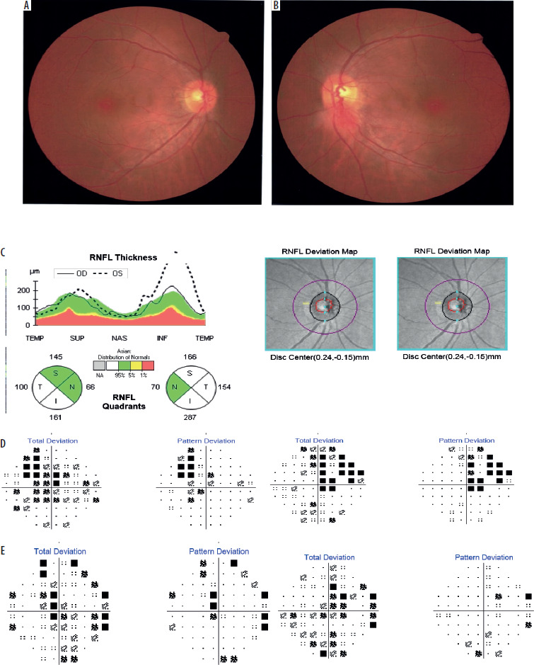
A 46-year-old woman weighing 62 kg presented with a three-week history of progressive painless bilateral visual loss. She had been undergoing treatment for pulmonary TB with a standard four-drug regimen consisting of isoniazid, rifampicin, pyrazinamide, and ethambutol (administered at a dose of 1500 mg/day or 25 mg/kg/day) for the past six months. The patient had completed anti-TB therapy two days prior to her initial ophthalmological evaluation. The patient’s medical history was unremarkable. At presentation, her BCVA was 0.63 in the right eye (OD) and 0.8 in the left eye (OS). Colour vision assessment using Ishihara plates yielded scores of 8/38 for OD and 20/38 for OS, whereas the Farnsworth test revealed no abnormalities. Contrast sensitivity, evaluated using the Mars contrast sensitivity chart, reduced to 1.25% in both eyes. Fundus examination revealed a normal right optic disc (Figure 1A) and left inferior optic disc swelling and peripapillary haemorrhage (Figure 1B). OCT revealed left thickening of the inferior RNFL (Figure 1C). Visual field testing using automated Humphrey perimetry indicated a bitemporal hemianopic defect (Figure 1D). CT findings were unremarkable. The patient was initiated on a regimen of Q10 coenzyme (90 mg), methylcobalamin (500 mcg), and zinc (20 mg) daily. At the one-month follow-up, her BCVA remained at 0.63 in both eyes; however, improvements were observed in both colour vision and contrast sensitivity. Visual field assessment also revealed some improvements (Figure 1E). At the five-month follow-up, her BCVA had improved to 1.0 in both eyes, with normalization of colour vision and contrast sensitivity.
Figure 1
Results of ophthalmologic analyses in Patient 1. A) Fundus photograph of the right eye at the initial visit. B) Fundus photographs of the left eye showing inferior swelling with peripapillary haemorrhage. C) Optical coherence tomography scan findings at the initial visit revealing thickening of the inferior retinal nerve fibre layer (RNFL) in the left eye. D) Automated perimetry results at the initial visit showing a bitemporal hemianopia defect. E) One-month follow-up after discontinuation of ethambutol. Note that the visual field defect was ameliorated (compared with the results at initial examination)
Patient 2
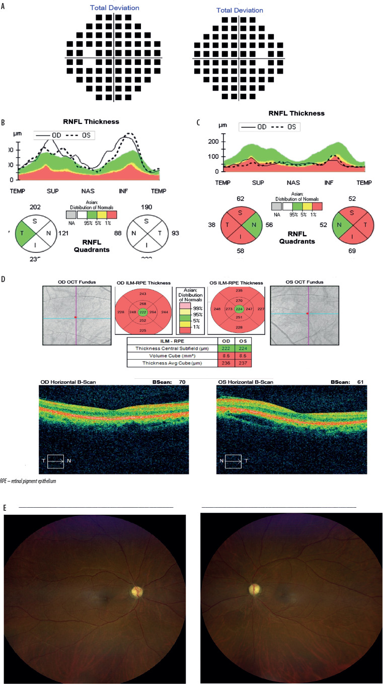
A 45-year-old woman (weight, 54 kg) presented with a two-week history of progressive bilateral visual loss. She had been receiving a TB regimen with ethambutol at a dose of 1000 mg/day (18.5 mg/kg/day) for five months. Her medical history revealed systemic hypertension but no prior ocular disease. At presentation, the BCVA was 0.2 OD and 0.125 OS. Colour vision was markedly reduced (1/38 OD and 0/38 OS), and contrast sensitivity was severely impaired (> 25% in both eyes). Bilateral optic disc swelling was observed, and visual field testing revealed complete field loss (Figure 2A). OCT revealed bilateral thickening of the superior and inferior RNFL (Figure 2B). CT findings were unremarkable. The patient was administered the same treatment as Patient 1 along with 10 mg amlodipine. Despite treatment, the BCVA declined to 1/60 in both eyes within two weeks. Ethambutol was discontinued one month after symptom onset, and the remainder of the anti-TB regimen was completed over a duration of nine months. Follow-up OCT revealed thinning of the RNFL (Figure 2C) and macula (Figure 2D). At the one-year follow-up, the BCVA remained at 1/60 in both eyes, with persistent colour vision and contrast deficits. Fundus examination revealed bilateral optic atrophy.
Figure 2
Results of ophthalmologic analyses in Patient 2. A) Automated perimetry results at initial visit, revealing complete loss of visual field. B) Optical coherence tomography (OCT) scan findings at initial visit, revealing increased thickness of the superior and inferior retinal nerve fibre layers (RNFL) in both eyes. C) OCT scan findings at nine-month follow-up, revealing decreased thickness of the RNFL in both eyes. D) Macular OCT scan findings at nine-month follow-up, revealing decreased thickness of the internal limiting membrane (ILM) in both eyes E) Fundus examination after one-year follow-up, showing optic atrophy both eyes
Patient 3
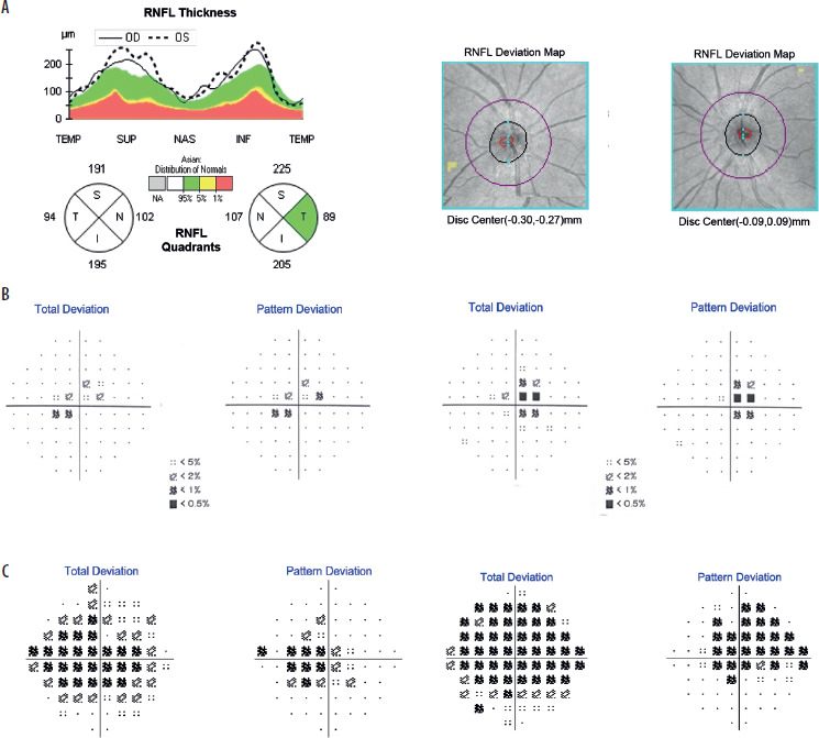
A 33-year-old man (weight, 69 kg) presented with a one-month history of progressive bilateral vision loss. He had been receiving ethambutol at a dose of 1500 mg/day (21 mg/ kg/day) for eight months. His medical history included diabetes mellitus, which was managed with metformin. At presentation, the BCVA was 3/60 OD and 0.2 OS. Colour vision was impaired (4/38 OD and 7/38 OS using the Ishihara plates), although Farnsworth testing produced normal findings. Contrast sensitivity was reduced to 25% OD and 1.25% OS. Fundus examination revealed swelling of the bilateral superior and inferior optic discs. OCT revealed thickening of the inferior RNFL in both eyes (Figure 3A), and visual field testing revealed bilateral central scotomas (Figure 3B). CT findings were unremarkable. Ethambutol treatment was discontinued, and the patient was started on the same regimen as the other patients. Two weeks later, the BCVA had improved to 0.2 in both eyes, but the central scotomas had enlarged (Figure 3C). However, there was partial recovery in colour vision and contrast sensitivity. By two months, the BCVA had improved to 0.63 OD and 0.5 OS, with restoration of other visual functions.
Figure 3
Results of ophthalmologic analyses in Patient 3. A) Optical coherence tomography scan findings at initial visit showing increased thickness of the superior and inferior retinal nerve fibre layers (RNFL) in both eyes. B) Automated perimetry results at initial visit, showing central scotoma in both eyes. C) Automated perimetry results at two-week follow-up, showing increased central scotoma in both eyes
Patient 4
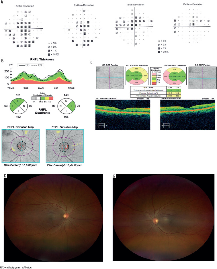
A 46-year-old woman (weight, 41 kg) presented with a three-month history of gradual, painless bilateral visual loss. The patient had been undergoing treatment for multi-drug-resistant pulmonary TB for 14 months. For the past 7 months, her treatment has included cycloserine (250 mg/ day), pyrazinamide (1500 mg/day), and ethambutol (1000 mg/day, < 25 mg/kg/day). One month before the presentation, ethambutol was discontinued and replaced with levofloxacin (750 mg/day). The patient’s medical history was unremarkable. Initial examination revealed a BCVA of 2/60 OD and 0.05 OS, severely reduced colour vision (1/38 in both eyes using the Ishihara plates), and contrast sensitivity of 2.5% bilaterally. Fundoscopy revealed bilateral hyperaemic optic discs with blurred margins. Visual field testing revealed bilateral central scotomas (Figure 4A). OCT revealed thickening of the superior and inferior RNFL (Figure 4B); however, macular OCT showed that the internal limiting membrane was intact (Figure 4C). CT findings were unremarkable. The patient was treated using the same regimen as the other patients. At one month, her BCVA improved to 0.5 in both eyes, with colour vision improving to 14/38. By the two-month follow-up, BCVA returned to 1.0 in both eyes, with full recovery of visual function and normal fundus appearance (Figures 4D and 4E).
Figure 4
Results of ophthalmologic analyses in Patient 4. A) Automated perimetry results at initial visit, showing central scotoma in both eyes. B) Optical coherence tomography (OCT) scan findings three-weeks after initial visit, showing increased thickness of the superior and inferior nasal retinal nerve fibre layers (RNFL). C) Macular OCT scan findings three-weeks after initial visit, showing a normal internal limiting membrane (ILM) layer. D) Fundus examination at follow-up in the right eye, revealing with slight blurry margins of the superior and inferior optic nerves. E) Fundus examination at follow-up in the left eye, revealing slight blurry margins of the superior and inferior optic nerves
DISCUSSION
Clinical presentation
Herein, we report four cases of ETON (listed in Table I), a condition associated with drug toxicity that typically manifests as progressive, bilateral, and painless vision impairment. This form of central vision loss is generally symmetric; however, there might be instances where patients initially manifest asymmetrical or sequential vision defects [7]. All the patients described in this series experienced bilateral, painless visual loss, with symptom durations ranging from two to 12 weeks prior to their initial ophthalmic examination. Each patient had a history of ethambutol use as part of their TB treatment. Dyschromatopsia is a common finding in toxic optic neuropathies and is usually detected using Ishihara, Farnsworth, or Panel D-15 tests [8]. In our case series, all patients exhibited impaired colour vision when tested using the Ishihara plates, while the results of the Farnsworth test were within normal limits. Early ETON commonly presents with red-green colour discrimination deficits, making the Ishihara test particularly useful for its early detection [9]. Different studies of ETON have reported varied patterns of visual field defects. Common patterns include central or cecocentral scotomas, peripheral field constriction, or bitemporal hemianopia [6]. All four patients exhibited visual field defects; notably, Patient 1 had bitemporal hemianopia, which may be attributed to ethambutol-induced damage to the nasal crossing fibres in the optic chiasm [5].
Table I
Summary of clinical characteristics, management, and outcomes
FUNDUS AND IMAGING FINDINGS
Fundus examination may initially appear normal, although signs such as optic disc hyperaemia or small peripapillary haemorrhage can sometimes be observed. In the early stages, toxic optic neuropathy may show minimal clinical change on fundoscopy; however, delayed diagnosis and intervention can lead to optic disc pallor, typically beginning at the temporal margin [2, 9, 10]. Fundoscopy findings among our patients revealed a range of features, including normal-appearing discs, disc swelling, peripapillary haemorrhage, and optic atrophy. RNFL analysis using OCT scans typically shows thinning in the later stages of ETON [6]. However, other studies have reported RNFL thickening during the early phases of toxicity [11]. The initial OCT scans of our patients showed RNFL thickening, predominantly in the inferior regions, with relative thinning of the temporal regions.
Because magnetic resonance imaging was not available at our institution, brain imaging was performed using CT scans, the findings of which were unremarkable in all cases. Imaging tests are important to rule out compressive optic neuropathy, because in cases of toxic aetiology, the findings look normal [9].
TREATMENT AND OUTCOMES
Each patient was prescribed ethambutol for durations ranging from 5 to 8 months. These durations exceeded those outlined in the national TB treatment protocol, which recommends a 4-month course of isoniazid (5 mg/kg/day), rifampicin (10 mg/kg/day), pyrazinamide (25 mg/kg/day), and ethambutol (15 mg/kg/day), followed by 2 months of isoniazid and rifampicin for new adult cases [12]. In our patients, the prolonged use of ethambutol was probably necessitated by treatment failure, persistent infection, or the need for second-line regimens for managing multi-drug-resistant TB. Prolonged ethambutol exposure increases the likelihood of toxicity, especially when dosages exceed 15 mg/kg/ day. Toxicity is more commonly reported at doses ≥ 25 mg/ kg/day [9], and all of the four patients received ethambutol doses > 15 mg/kg/day. The mechanism potentially underlying ethambutol-induced optic neuropathy involves the inhibition of arabinosyltransferase, an enzyme essential for mycobacterial cell wall synthesis. This inhibition is believed to disrupt mitochondrial oxidative phosphorylation, increase the production of reactive oxygen species, and trigger retinal ganglion cell damage and apoptosis [10].
No specific therapy currently exists for ETON; therefore, immediate discontinuation of ethambutol is recommended when toxicity is suspected [13]. In all four patients, ethambutol treatment was stopped at either the time of diagnosis or shortly thereafter. Supportive treatment with coenzyme Q10 (90 mg/ day), methylcobalamin (500 mcg/day), and zinc (20 mg/day) was initiated. Coenzyme Q10 has demonstrated protective effects on retinal ganglion cells in animal models [14], while zinc may counteract the chelating effects [9]. Supplementation with B vitamins, especially B12, is commonly used to manage mitochondrial optic neuropathies and is potentially beneficial in ETON owing to shared pathogenic mechanisms [10]. In our case series, three of the four patients (75%) showed some degree of visual improvement after discontinuation of ethambutol treatment and initiation of adjunctive therapy.
RISK FACTORS AND PROGNOSIS
Several risk factors are known to potentiate neurotoxic effects. These include higher dosage, extended therapy duration, systemic hypertension, alcohol or tobacco use, immunosuppression, and concurrent HIV infection [9]. Despite similar management protocols, visual recovery varied in our patients. Patients 1 and 4 experienced complete recovery of vision, Patient 3 showed partial improvement, and Patient 2 progressed to bilateral optic atrophy. Interestingly, despite Patient 2 exhibiting relatively preserved visual acuity, the eventual outcome was poor. This contrasts with the results in Patients 1 and 4, who had worse initial acuities but exhibited significant recovery. A previous study indicated that patients with an initial visual acuity worse than 6/60 tended to have unfavourable outcomes. However, this finding was not universally applicable in our patient. Additionally, Patient 2 had a history of systemic hypertension, which may have adversely affected the prognosis. Comorbid conditions such as advanced age, hypertension, and renal insufficiency have been associated with poorer outcomes in ETON [15].
CONCLUSIONS
This case series highlights the variable clinical presentations and visual outcomes of ETON. Although early identification and cessation of ethambutol treatment, concordant with supportive therapy using coenzyme Q10, methylcobalamin, and zinc, appeared to be beneficial in most patients, the prognosis remained inconsistent. Three of the four patients experienced some degree of visual recovery, and one progressed to optic atrophy despite timely management.
These findings emphasise that initial visual acuity may not always predict visual outcomes and that comorbidities such as hypertension could contribute to a poorer prognosis. Given the risk of irreversible visual impairment, regular ophthalmo-logical monitoring is critical for patients receiving ethambutol therapy, particularly those on high-dose or long-term treatment regimens.