INTRODUCTION
The constant technological progress and better quality of new intraocular lens (IOL) models may enhance the functional outcomes of cataract surgery and, consequently, patients’ quality of life. Monofocal implants remain the most widely used type of IOLs and provide good visual acuity (VA) at a single focal distance.
The first artificial IOLs were designed with monofocal optics, offering correction for a fixed distance, usually for far distance. Consequently, patients required spectacle correction for intermediate and near tasks after surgery. The only possibility to achieve spectacle independence is to achieve functional VA at distance, intermediate, and near ranges.
In order to address this problem, as well as to fulfill patients’ expectation of good vision for various distances, many new types of IOLs were designed, including: extended depth-of-focus (EDoF), bifocal (Bi), trifocal, multifocal (MIOL), and accommodating lenses [1, 2].
A defocus curve (DC) is used to assess the range of expected binocular VA after bilateral implantation of a particular IOL model [3]. To create a DC, binocular VA should be measured at a fixed distance. After placing in front of the patient minus spherical lenses of progressively increasing power, usually in half diopter (D) steps, VA is recorded. A defocus step of −1.50 D represents intermediate vision at a distance of 66 cm, while −2.50 D represents near vision at a distance of 40 cm [3].
The aim of this study was to evaluate the DC (also known as a depth of field curve) in patients after bilateral cataract extraction using phacoemulsification, who had an artificial IOL implanted in both eyes: an EDoF, MIOL, Bi, or a standard monofocal acrylic foldable IOL.
MATERIAL AND METHODS
This was a single-site, prospective, randomized, controlled trial. The study was approved by the Bioethics Committee of the Medical University of Lodz (Approval number RNN/ 109/15KE).
This research followed the tenets of the Declaration of Helsinki, and after explaining the nature and possible consequences of the study, informed consent was obtained from all patients. The study group consisted of 100 patients (79 women and 21 men), mean age 71 ±8 years, who underwent phacoemulsification with binocular IOL implantation or various lenses, who met the inclusion criteria and underwent surgery at the Department of Ophthalmology, Medical University of Lodz, Poland. After providing consent to participate in the study, patients were randomly assigned to one of four study groups:
group 1 – EDoF Tecnis Symfony (Johnson & Johnson, USA) (n = 20), age 72 ±6 years,
group 2 – MIOL Diffractiva-aA (HumanOptics, Germany) (n = 30), age 73 ±7 years,
group 3 – Diffractiva-aA Bi (HumanOptics, Germany) (n = 25), age 69 ±7 years,
group 4 – monofocal Aspira-aA lens (HumanOptics, Germany) (n = 25), age 69 ±10 years.
The patients’ age did not differ significantly between the examined groups.
The lens models were selected based on various properties of the optical part and their availability.
The inclusion criteria were bilateral cataract in patients with preoperative best-corrected distance VA ranging from 0.3 to 0.8 (decimal) and with corneal astigmatism up to 0.75 D.
The exclusion criteria were concomitant eye diseases (e.g. congenital anomalies of the eyeball, corneal diseases, scars, opacities, preoperative corneal endothelial density below 1500 cells/mm2, dry eye syndrome, glaucoma, retinal and macular diseases, previous ocular injury, amblyopia, strabismus, uveitis and a history of optic neuropathy, diabetic retinopathy, previous central retinal artery occlusion or central retinal vein occlusion, eyeball tumors), as well as the presence of other diseases that may affect postoperative VA (diabetes, hypertension, stroke, multiple sclerosis, connective tissue diseases). Professional drivers, pilots, and pregnant or lactating women were also excluded from the study.
Pre-operative management
Biometric calculations were performed using an IOL Master 700 device (Zeiss), and the target refraction was emmetropia.
All patients received topical antibiotic drops of levofloxacin (Oftaquix, Santen) four times daily for three days before the surgery.
All procedures were performed under local anesthesia – topical proxymetacaine drops (Alcaine, Alcon) and lidocaine gel (2%) (Lignocainum, Jelfa), as well as intracameral anesthesia with 1% lidocaine solution (Lignocainum hydrochloricum WZF, Polpharma). The surgical procedures were performed by three experienced surgeons.
In all cases, the pupils were dilated before surgery using a 1% solution of tropicamide (Tropicamidum WZF, Polfa) and a 10% solution of phenylephrine (Neosynephrin-POS, Ursapharm).
Surgical technique
Phacoemulsification was performed through a 2.2 mm clear corneal self-sealing temporal incision. After entering the anterior chamber and administering the ophthalmic viscosurgical device (OVD), continuous circular capsulorhexis was performed with microforceps, then two paracenteses were created with an MVR 20G knife. An infusion fluid (balanced salt solution) was used to perform hydrodissection. Phacoemulsification was performed using the Stellaris system (Bausch & Lomb). Cortical masses were removed by irrigation and aspiration. Then, after injecting the OVD into the anterior chamber, the appropriate IOL for a given group was implanted using an injector. The IOL was inserted in group 1 using the Johnson & Johnson Unfolder Platinum 1 Series delivery system injector, and in groups 2, 3, and 4 using the Medicel Accuject/Naviject injector. After removal of the OVD, the corneal wounds were sealed with 0.9% saline (NaCl) solution and left unsutured.
Postoperative management
After the surgery, all patients used steroid drops with antibiotics containing 3 mg/ml of tobramycin with 1mg/ml of dexamethasone (Tobradex, Alcon), four times daily for three weeks, then twice daily for one week.
Examinations
All patients were examined before the surgery and 1 month and 6 months postoperatively. The second eye was operated on 4–6 weeks after the first one. Preoperative examination included a full ophthalmic examination, assessment of cataract in the LOCS III system, biometry, and all the examinations conducted postoperatively. Postoperative examinations included a full ophthalmic examination, assessment of uncorrected and best corrected VA for distance, intermediate (60 cm), and near (40 cm) vision, as well as the DC.
Statistical analysis
Statistical analysis was performed using parametric tests. Differences between groups were assessed using one-way analysis of variance (ANOVA) with Tukey’s post-hoc test, with a 95% confidence interval. All calculations were performed using Microsoft Excel and XLSTAT (Addinsoft, 2008). A p-value < 0.05 was considered statistically significant.
RESULTS
The DC was assessed at 1 and 6 months after cataract surgery in both eyes (Tables I and II). In all groups the results of DC examined 1 month and 6 months postoperatively were similar.
Table I
Comparison of the results of the bilateral defocus curve 6 months after cataract surgery [visual acuity (VA), Snellen: mean ± standard deviation]. Blue background represents good vision (VA ≥ 0.8), grey background represents acceptable vision (VA = 0.6–0.8)
Bi – bifocal; D – diopters; EDoF – extended depth of focus; IOL – intraocular lens; MIOL – multifocal IOL Bold values indicate the highest mean VA achieved among the tested IOL groups.
Table II
Statistical significance of differences in the bilateral defocus curve (DC) between the examined groups at 6 months after cataract surgery in both eyes – for each power [from –5 diopters (D) to +1 D]. Values represent p-values for intergroup differences at each defocus level
Binocular VA was assessed using trial lenses of varying optical power (from –5.00 D to +1.00 D, in 0.5 D steps) to simulate different viewing distances. Results were presented in the decimal Snellen format. Two thresholds were defined: 0.8 (good vision) and 0.6 (acceptable vision).
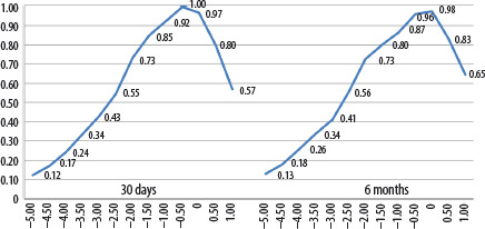
Six months after the second eye surgery, the DC in group 1 (EDoF Tecnis Symfony ZXR00 IOL) demonstrated a continuous range of good visual acuity from (VA ≥ 0.8) from −1.50 to +0.50 D. Acceptable visual acuity (VA ≥ 0.6) extended from −2.00 to +1.00 D. The curve showed a monophasic profile (without gaps in the intermediate range) corresponding to a single focus extending from intermediate to far distances (50 cm to infinity) (Figure 1).
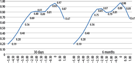
In patients from group 2 (Diffractiva-aA MIOLs), good visual acuity was achieved over a defocus range from –2.50 D to +0.50 D, while acceptable vision ranged from –3.00 D to +1.00 D. The DC profile biphasic, with one peak corresponding to near and intermediate vision, and a second peak corresponding to distance VA (33 cm to infinity) (Figure 2).
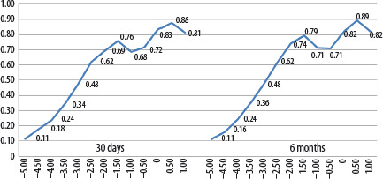
Good vision in group 3 (Diffractiva-aA Bi IOLs) ranged from 0 to +1.00 D, as well as –1.50 D, and acceptable vision ranged from –2.50 D to –0.50 D. The DC curve was biphasic, with peaks corresponding to near focus (40 cm) and distance (infinity) (Figure 3).
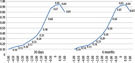
In group 4 (Aspira-aA monofocal IOLs), good vision (≥ 0.8) was achieved over a defocus range from –0.50 D to +1.00 D, while acceptable vision was achieved at –1.00 D. The DC showed a single peak corresponding to distance vision (from 1 m to infinity) (Figure 4).
In our study, it was found that the best results of binocular distance vision (0 D, infinity) 6 months after cataract surgery were obtained in eyes with an EDoF IOL (0.98), MIOL (0.98), and a monofocal IOL (0.93), and they were significantly worse in eyes with a Bi IOL (0.82) (Figure 5).
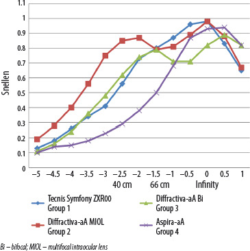
In addition, there was a significant difference at +0.5 D between a monofocal IOL (0.94), a Bi IOL (0.89), and an EDoF IOL (0.83). However, at +1.0 D, monofocal IOLs (0.82) were significantly different from Bi IOLs (0.82), MIOL (0.67), and EDoF IOL (0.65).
The results of binocular vision for intermediate distances (–1.5 D, 66 cm) offered by an MIOL, Bi, and EDoF IOL were comparable (0.8) and were significantly worse with a monofocal IOL (0.5).
At near distance (–2.5D, 40 cm) binocular visual acuity was highest with MIOLs (0.85), followed by Bi (0.62), EDoF (0.56), and monofocal IOL (0.29). Monofocal IOLs demonstrated the lowest near visual acuity among the study groups.
Similar results were observed at –3.00 D (33 cm), where satisfactory VA was achieved only in patients with MIOLs (0.75), while the remaining groups showed values below 0.5.
The EDoF IOL provided good (≥ 0.8) or acceptable (≥ 0.6) binocular vision over a defocus range from –2.00 D to +0.50 D (from 50 cm to infinity). The DC showed a monophasic profile with a single extended focus from intermediate distances (50–100 cm with acceptable VA at 50 cm, good at 66–100 cm) to far distance (> 2 m, good VA).
MIOLs provided good or acceptable vision over a defocus range from –3.00 D to +1.00 D (from 33 cm to infinity). The curve profile was biphasic, with one peak corresponding to good vision for near (33–40 cm) and intermediate distance (50–100 cm), while the second peak corresponded to good vision for distance vision (> 2 m).
Bi IOLs provided good or acceptable vision over a defocus range from –2.50 D to +1.00 D (40 cm to infinity), with a biphasic DC having two peaks: acceptable near and intermediate distance (40–100 cm) and a good distance VA (to infinity).
Monofocal IOLs provided good or acceptable binocular vision over a defocus range from –1.00 D to +1.00 D (from 1 m to infinity), which produced a monophasic DC with a single clear peak for distance vision (> 2 m), as well as acceptable vision from a distance of 1 m.
One month after cataract surgery, significant differences in binocular visual acuity were observed between the study groups across multiple defocus levels (Table II). Overall, group 2 (MIOLs) demonstrated superior visual acuity at near and intermediate defocus levels compared with the other groups, whereas group 4 (monofocal IOLs) achieved better visual acuity at positive defocus levels. Detailed pairwise comparisons are presented in Table II.
DISCUSSION
It is known that various types of IOLs have different optical properties, so choosing an appropriate lens to fulfill individual needs is important.
Evaluating the DC is especially important when comparing different types of IOLs, as it defines binocular visual performance of patients at various distances.
Gil et al. [4] assessed the results of phacoemulsification with implantation of 6 different lenses: EDoF Tecnis Symfony ZXR00 (n = 20), Tecnis MF [ZKB00 (n = 20), ZLB00 (n = 20)], AcrySof ReSTOR SV25T0 MF (n = 19), AT LISA 809M (n = 18), AT LISA tri 839MP (n = 19). The authors found that the Tecnis Symfony ZXR00 IOL provides a defocus range from –1.50 D to +0.50 D (66 cm to infinity) without any curve fading, achieving a distance and intermediate distance better than 0.05 logMAR (corresponding to > 0.8 decimal) [4]. These results are identical to our study.
Pedrotti et al. [5] compared the results of implantation of 4 different lenses: EDoF Tecnis Symfony ZXR00 (n = 55), ReSTOR +2.5 and +3.0 (n = 50), and a monofocal Tecnis IOL (n = 30). They found that patients with the Tecnis Symfony ZXR00 IOL achieved good vision in the defocus range from –1.5 D to 0 (66 cm – infinity), and acceptable – at –2.0 D (50 cm), with a smooth DC. In contrast, after implanting a monofocal lens, which provides a defocus range from 0 to –0.50 D (infinity), patients obtained a good or acceptable VA at –1.00 D (1 m). Significantly better binocular VA was found in patients with an EDoF IOL. These results were in accordance with our findings.
Hamid et al. [6] assessed the DC in the range from 0 D to –3.0 D 6 months after phacoemulsification with implantation of three different lenses: trifocal diffractive IOL AT LISA tri 839 MP (n = 50), PhysIOL FineVision (n = 50), and IOL EDoF Tecnis Symfony ZRX00 (n = 50). The obtained IOL Symfony defocus profile was the best, with the range of good distance and intermediate distance vision from 0 D to –1.50 D (66 cm to infinity). DC results for intermediate distances of an EDoF lens outperformed the rest. In contrast, near visual acuity was better in patients implanted with diffractive trifocal MIOLs. A characteristic second peak of the DC was observed between −2.0 and −3.0 D, corresponding to good near visual acuity with the FineVision lens, whereas the AT LISA lens provided lower visual acuity within this range.
Esteve-Taboada et al. [7] investigated the optical quality of three IOLs: Tecnis Symfony, AT LISA, and FineVision. The researchers found that the Tecnis Symfony EDoF IOL exhibited two main regions of good visual performance, corresponding to distance and intermediate vision. On the other hand, the diffractive lenses FineVision and AT LISA exhibited three focal points each, providing distance, intermediate, and near vision. In addition, the FineVision lens showed superior optical quality for distance, the Tecnis Symfony IOL for intermediate distances, and the AT LISA IOL for near [8].
Monaco et al. [9] analyzed the DC results in 60 patients 4 months after cataract surgery with binocular implantation of three different IOLs: EDoF Tecnis Symfony ZXR00 (n = 20), trifocal AcrySof IQ Panoptix TFNT00 (n = 20), and monofocal AcrySof SN60WF (n = 20). The results of the DC showed significantly better VA at intermediate distance (at –1.5D) and for near distance (–2.5 to –4.0D) in patients with the Panoptix IOL compared to patients with the Symfony and the SN60WF lenses. Patients with a single-focus IOL compared to Tecnis Symfony EDoF had significantly worse VA (from –1.0 D to –4.0 D). In our study, the results of the DC for intermediate distances were comparable for groups 1 and 2, while group 2 (with MIOL) exhibited better VA for near. Patients with a monofocal IOL had the worst VA results (from –1.0 D to –5.0 D).
Farvardin et al. [10] one year after cataract surgery with binocular implantation of AcrySof IQ PanOptix TNFT00 (Alcon) (n = 20) or Tecnis Symfony ZXR00 (AMO) lenses (n = 20) reported results for a binocular DC (–0.5 to 0.5 D) that were comparable for both lenses (0.10 to 0 logMAR). The DC for intermediate distances was slightly better in the PanOptix IOL group, but did not differ significantly from the Symfony IOL; however, this difference was significant for near distances.
Lubiński et al. [11], one year after cataract surgery in 40 patients with bilateral implantation of AT LISA tri 839 MP (n = 20) or Tecnis Symfony ZRX00 (n = 20) lenses, evaluated the binocular DC in the range from 2.0 D to –4.0 D. The obtained VA results in the DC for distance (2.0 D, 1.0 D, 0 D) and intermediate vision (–1.5 D, 66 cm) were similar for both groups, while the remaining parameters significantly (p < 0.05) differed in favor of AT LISA MIOL. Our results in group 2 are identical.
Dexl et al. [12] examined 24 patients 6 months after cataract surgery with a Diffractiva-aA MIOL and assessed the binocular DC. They observed two peaks of maximum VA: for distance (0 D) −0.01 ±0.05 logMAR (≈1.02 Snellen) and for near (−2.5 D) 0.03 ± 0.07 logMAR (≈0.9 Snellen). In addition, satisfactory vision was observed for intermediate distances (–1.5 D/66 cm): 0.14 ±0.08 logMAR (≈0.7 Snellen). These results are very similar to our results in group 2.
Schrecker et al. [13] one year after cataract surgery with Diffractiva-aA MIOL implantation assessed a monocular DC ranging from +1.0 D to –3.5 D every 0.5 D. The DC was Bi with a far point at 0 D (with VA 0.1 logMar/0.8 Snellen) and a near point at –2.50 D. The range of good intermediate vision ranged from –1.0 D to –2.0 D. The results of the abovementioned parameters in our group 2 are identical.
Similar results were also obtained by Grabner et al. [14], who compared the results of the DC 3 months after cataract surgery in patients (n = 30) with binocular Diffractiva-aA MIOL +3.5 D with a group of patients (n = 30) who received a diffractive AcrySof Restor +3.0 D MIOL in both eyes. DC for both lenses were biphasic, with the best VA for distance at 0 D and near distance (40 cm) at –2.5 D. However, patients with the Diffractiva-aA MIOL had significantly better VA (–1.50 D, ≈67 cm) for intermediate distances. Nevertheless, both IOLs provided a range of good vision for near and distance and comfortable vision at intermediate distances.
Rasp [15] presented 6-month results of binocular Diffractiva-aA MIOL implantation in 17 patients (34 eyes). Good intermediate vision was reflected by a monocular DC, with the lowest point (VA) at 0.20 logMAR (20/30 or 0.6 Snellen).
Lasta et al. [16] assessed the DC 3 months after cataract surgery in 60 patients with the monofocal Aspira-aA or the Tecnis ZCB00 IOL. The corresponding values were 0.61 ±0.33, and 0.65 ±0.29, respectively. In our study the DC in group 4 was comparable (0.60).
Schrecker et al. [17] assessed the DC (from 2.00 D to –3.00 D every 0.50 D) in patients after cataract surgery with a standard aspherical aberration-free IOL (Aspira-aA, n = 28) or IOL with personalized spherical aberration correction (Invidua-aA, n = 57). Three months after the procedure, the researchers found that both IOLs provided good or acceptable vision in the defocus range from –1.00 D to +1.00 D (from 1 m to infinity), which produced a single-phase DC with a single distinct peak for distance (> 2 m) with acceptable vision from a distance of 1 m. Our results for the Aspira-aA lens were identical.
In summary, patients differ in age, lifestyles, and visual requirements. Therefore, selection of the most appropriate lens should be individualized and take into account not only a single property of the IOL, such as the DC, but many additional factors, including the optical properties of the lens, the VA achieved at different distances, contrast sensitivity, and unwanted optical phenomena. Patient-related factors that should be considered when selecting an IOL include lifestyle, visual goals, presence of astigmatism, age, desire for spectacle independence (e.g., reading, computer work, driving), hobbies, occupation, night driving frequency, general health, and tolerance of potential side effects, e.g. the risk of glare, halos, or reduced contrast. Due to the complexity of these multiple factors, IOL selection should be individualized to fulfill the needs of a particular patient.
The main limitations of this study were the relatively small sample size and the short follow-up period (6 months), which may have influenced the results.
CONCLUSIONS
The DC showed that comparably good binocular VA for distance was obtained in eyes with EDoF lenses, diffractive- refractive MIOLs and monofocal lenses, while it was significantly worse in eyes with Bi IOLs. The DC showed that binocular VA for intermediate distances was comparably good for EDoF and MIOLs, slightly worse for Bi IOLs and significantly worse for monofocal lenses. Binocular near VA was the highest with MIOLs, slightly lower with EDoF and Bi IOLs, and the lowest with monofocal lenses. Given the different optical properties of various IOLs and patients’ different expectations, the IOL should be individually selected to fulfill the needs of a particular patient.