twitter
en POLSKI
eISSN: 2719-3209
ISSN: 0023-2157
Klinika Oczna / Acta Ophthalmologica Polonica
Current issue Archive Videos Articles in press About the journal Supplements Editorial board Reviewers Abstracting and indexing Subscription Contact Instructions for authors Ethical standards and procedures
Editorial System
Submit your Manuscript
SCImago Journal & Country Rank
Share:
Share:
Case report

Syphilitic optic neuritis and uveitis – case report

Karolina Bonińska
1, 2, 3
,
Hanna Sobutka
1
,
Dagmara Mrowicka
1
,
Michał Post
3, 4
,
Tomasz Dybek
1
,
Maciej Bednarski
1, 2
,
Sławomir Cisiecki
1, 2

1.
Ophthalmology Department, Dr. K. Jonscher Municipal Medical Center in Lodz, Poland
2.
Julianów Medical Center in Lodz, Poland
3.
Club 40, Polish Society of Ophthalmology, Poland
4.
Department of Ophthalmology, Independent Public Clinical Ophthalmology Hospital, Medical University of Warsaw, Warsaw, Poland
KLINIKA OCZNA 2023, 125
Online publish date: 2023/12/09
Article files
- KO-00459_EN.pdf  [2.18 MB]
- KO-00459_PL.pdf  [2.18 MB]
Get citation
 
PlumX metrics:
 

INTRODUCTION

Syphilis is a sexually transmitted infectious disease caused by the bacterium Treponema pallidum. The primary modes of transmission include sexual contact with an infected person and vertical transmission. Syphilis is a chronic multisystem disease characterized by a wide spectrum of symptoms and following a cyclical course with alternating periods of exacerbation and latency. Involvement of the nervous system, including optic neuritis and other ocular manifestations, can occur at any stage of syphilis infection.

Case report

A 34-year-old male patient was referred to our Department due to uveitis in the right eye and optic neuritis in the left eye. The patient denied any comorbidities and chronic medication use.
Based on the physical examination and the medical records provided, it was determined that the onset of ophthalmic symptoms had occurred four months earlier. At that time, the patient was hospitalized at another ophthalmology center because of painless deterioration of vision in the right eye to the level of hand movement in front of the eye, which had persisted for two days. The patient reported no complaints in the left eye. Despite conducting examinations, the underlying cause of progressive vision loss in the right eye remained unidentified. Intravenous methylprednisolone treatment was initiated.
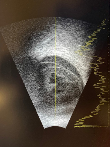
On the day of admission to our Department, the patient’s physical examination revealed loss of light perception in the right eye. The visual acuity in the left eye was 0.8. The intraocular pressure was 23 mmHg and 18 mmHg, respectively. Slit-lamp examination revealed, in the right eye, deposits on the corneal endothelium, single inflammatory cells in the anterior chamber, a non-reactive, unrounded pupil with posterior synechiae, and no fundus view. Ultrasound examination showed the presence of multiple densities in the vitreous and thickening of the choroid (Figure 1).
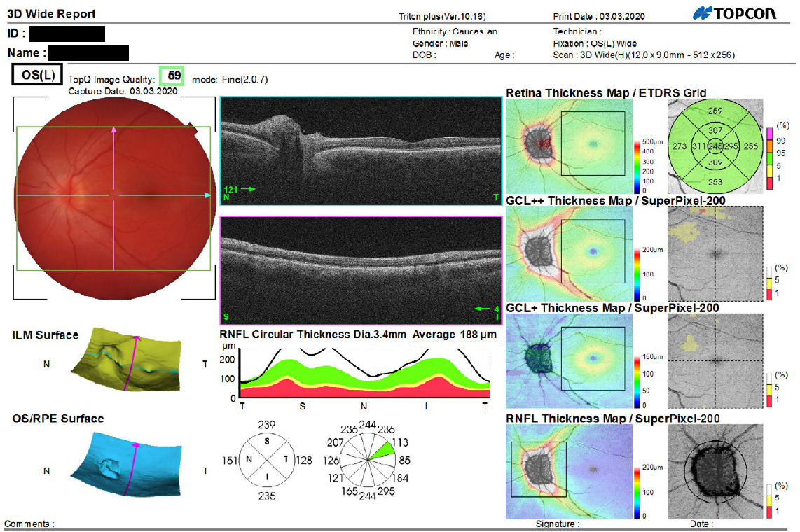
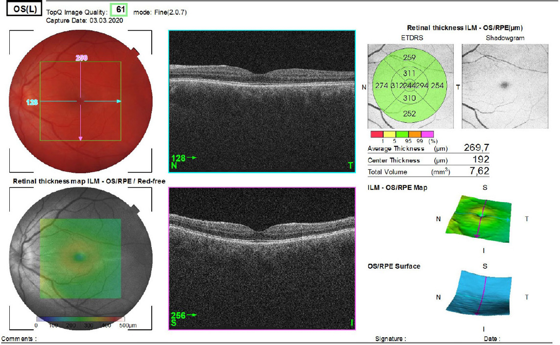
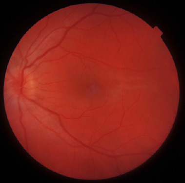
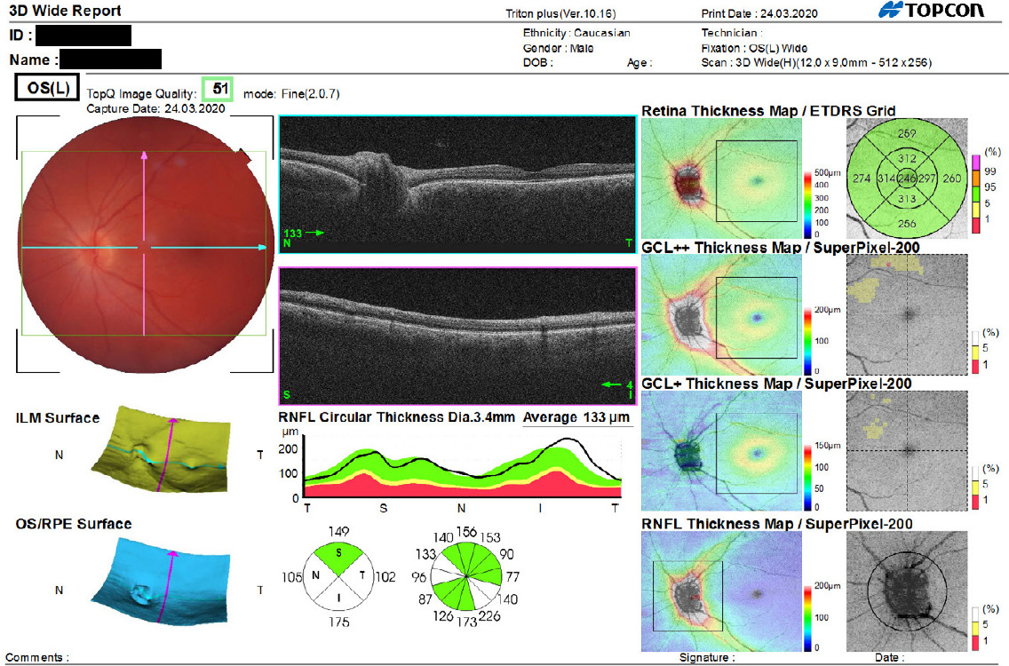
In the left eye, no pathologies were found in the anterior segment, but optic disc edema was observed (Figure 2). In addition, the patient’s optic nerve was assessed by spectral optical coherence tomography (SOCT) ((Figure 3). SOCT scan of the left macula was within the normal range (Figure 4). 
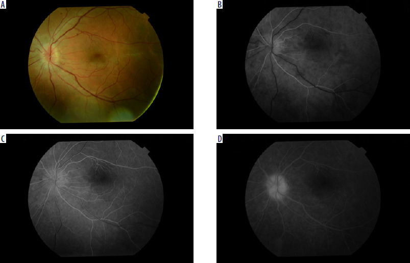
Fluorescein angiography showed multiple foci of diffuse hyperfluorescence with signs of late-phase leakage (Figure 5 A-D).
In the biochemistry panel, a number of abnormalities were found, including ESR (30 mm/h), doubtful IgM antibody test and positive IgG antibody test for Borrelia burgdorferii and HSV, and reactive WR-RPR result. Western blot assay for Lyme disease along with TPHA and FTA-ABS tests for syphilis were ordered for further verification.
The results of other laboratory tests (Quantiferon-TB, ANA, ANCA, RF, antibodies to Toxoplasma gondii, Toxocara spp., Bartonella henselae, HIV, HCV, HBV) were negative.
Contrast-enhanced MRI of the brain revealed an incidental finding – an arachnoid cyst in the right posterior cranial fossa, measuring 76 × 54 × 33 mm, deforming the right cerebellar hemisphere. Lung X-ray and abdominal ultrasound findings were normal. Neurological consultation revealed bilaterally positive Sterling’s and Rossolimo’s signs, and Babinski’s sign tendency on the left side.
During the patient’s stay in the Department, topical agents (dexamethasone drops, mydriatics) were applied to both eyes. In addition, the patient received intravenous steroid therapy (methylprednisolone at a dose of 1 g/day for a total of 5 days). A decrease in visual acuity to 0.5 was noted in the left eye.
After one week, on the day of discharge, the best-corrected visual acuity in the left eye was 0.8. The patient’s intraocular pressure was within the normal range. Physical examination revealed a raised optic disc with blurred margins (Figures 6, 7). Steroid therapy was maintained in the oral form (prednisone).
After receiving the examination results confirming the diagnosis of syphilis, oral antibiotic therapy (doxycycline 200 mg) was initiated. The patient was referred to the Neurology Outpatient Clinic to determine eligibility for lumbar puncture and potential continuation of treatment following the protocol for neurosyphilis. In addition, the patient was advised to schedule consultations at the Infectious Diseases Outpatient Clinic and the Dermatology Outpatient Clinic.
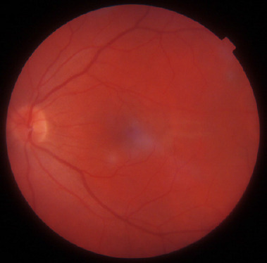
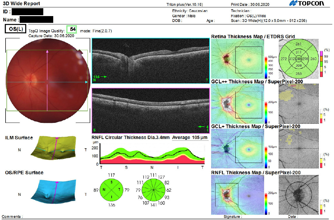
The patient remains under the care of ophthalmology and neurology specialists. The last examination, six months after hospitalization in the Ophthalmology Department, showed no sense of light in the right eye and visual acuity of the left eye at 1.0. Physical examination of the right eye revealed a clear cornea, clean anterior chamber, unrounded pupil, posterior synechiae, and pigment deposits on the lens. No abnormalities are found in the anterior segment and fundus of the left eye (Figure 8). The findings of the follow-up SOCT scan of the optic nerve were normal (Figure 9).

Discussion

The incidence of syphilis worldwide is rising, especially among high-risk populations. Those most at risk include homosexual men and HIV-positive individuals [1, 2]. Ocular manifestations are rare, found in only 1-5% of neurosyphilis cases [3]. They can occur at any stage of the disease, but are most common (4.6%) in secondary syphilis [4]. Approximately 75% of patients are male [2]. The mean age of onset of ocular symptoms is 48.6 years [5].
Syphilis can involve all ocular structures, but the most common manifestation is uveitis. Panuveitis occurs in 41% of patients with ocular syphilis, while optic nerve is involved in 22% [6]. The most common symptoms reported by patients include reduced visual acuity, ocular pain and headache, photophobia, red eyes, and increased lacrimation. In some cases, the disease may progress asymptomatically [3, 7-10]. Possible complications include the development of cataract, glaucoma, epiretinal membrane, optic nerve atrophy, or retinal detachment [8].
The first-line drug in the treatment of syphilis is crystalline penicillin at a dose of 18-24 million units/day for 10-14 days. An alternative is oral doxycycline at a dose of 200 mg twice a day for two to four weeks [1, 4].

Conclusions

Given the increased incidence of syphilis in recent years and the fact that ocular manifestations of the condition can mimic disease entities of various etiologies, it is important to include syphilis in the differential diagnostic process. In view of the broad range of symptoms, potential multiple organ involvement and associated diagnostic challenges, syphilis remains a disease requiring close collaboration of medical professionals of various specialties.

Disclosure

The authors declare no conflict of interest.
References

WPROWADZENIE

Kiła (syphilis) należy do chorób zakaźnych przenoszonych drogą płciową. Wywoływana jest przez bakterię, krętka bladego (Treponema pallidum). Najczęstszą drogą zakażenia jest kontakt seksualny z osobą chorą oraz droga wertykalna. Tę przewlekłą, wieloukładową chorobę cechuje szerokie spektrum objawów oraz fazowy przebieg z okresami zaostrzeń i utajenia. Zajęcie układu nerwowego, w tym zapalenie nerwu wzrokowego oraz inne objawy okulistyczne, może wystąpić na każdym etapie zakażenia kiłą.

Opis przypadku

Pacjent, lat 34, został skierowany na Oddziału Okulistyczny Miejskiego Centrum Medycznego im. dr. K. Jonschera z powodu zapalenia błony naczyniowej oka prawego oraz zapalenia nerwu wzrokowego oka lewego. Choroby współistniejące negował; żadnych leków na stałe nie przyjmował.
Na podstawie badania przedmiotowego oraz dostarczonej dokumentacji medycznej ustalono, że początek objawów okulistycznych miał miejsce 4 miesiące wcześniej. Chory był wówczas hospitalizowany w innym ośrodku okulistycznym z powodu bezbolesnego, występującego od 2 dni, pogorszenia widzenia w oku prawym do poziomu ruchu ręki przed okiem. Bez dolegliwości ze strony oka lewego. Pomimo wykonanych badań nie odnaleziono przyczyny postępującej utraty widzenia oka prawego. Zastosowano wówczas dożylne leczenie metyloprednizolonem.
W dniu przyjęcia na Oddział Okulistyczny w badaniu przedmiotowym stwierdzono brak poczucia światła w oku prawym, ostrość wzroku w oku lewym wynosiła 0,8. Ciśnienie wewnątrzgałkowe wynosiło odpowiednio 23 mm Hg i 18 mm Hg. W badaniu w lampie szczelinowej stwierdzono w oku prawym osady na śródbłonku rogówki, pojedyncze komórki zapalne w komorze przedniej, niereaktywną, odokrągloną źrenicę wraz ze zrostami tylnymi oraz brak wglądu w dno oka. Wykonane badanie ultrasonograficzne ujawniło liczne zagęszczenia w ciele szklistym oraz pogrubienie naczyniówki (rycina 1).
W oku lewym nie stwierdzono zmian patologicznych w obrębie przedniego odcinka, zaobserwowano natomiast obrzęk tarczy nerwu wzrokowego (rycina 2). Wykonano również spektralną optyczną koherentną tomografię (spectroscopic optical coherence tomography – SOCT) nerwu wzrokowego (rycina 3). Wykonane SOCT plamki oka lewego w granicach normy (rycina 4).
W angiografii fluoresceinowej stwierdzono liczne ogniska rozsianej hiperfluorescencji z cechami przecieku w fazach późnych (rycina 5 A-D).
Spośród badań biochemicznych od normy odbiegały następujące wyniki: OB 30 mm/godz., wątpliwy wynik przeciw- ciał w klasie IgM, dodatni w klasie IgG przeciwko Borrelia burgdorferi i HSV oraz reaktywny WR-RPR. W celu dalszej weryfikacji zlecono test Western blot w kierunku boreliozy oraz odczyn TPHA i FTA-ABS w kierunku kiły.
Pozostałe wyniki badań laboratoryjnych, m.in.: Quantiferon-TB, ANA, ANCA, RF, przeciwciała przeciwko Toxoplasma gondii, Toxocara spp., Bartonella henselae, HIV, HCV, HBV okazały się ujemne.
Wykonany rezonans magnetyczny mózgowia z kontrastem ujawnił przypadkowe znalezisko – torbiel pajęczynówki tylnego dołu czaszki po stronie prawej wielkości 76 × 54 × × 33 mm modelującą prawą półkulę móżdżku. Badanie rentgenowskie płuc i ultrasonograficzne jamy brzusznej nie wykazały odchyleń od normy. W trakcie konsultacji neurologicznej stwierdzono obustronny objaw Sterlinga i Rossolimo, tendencje do objawu Babińskiego po stronie lewej.
W czasie pobytu pacjenta na Oddziale wdrożono leczenie miejscowe do obojgu oczu: krople z deksametazonem, mydriatyki, oraz dożylną steroidoterapię (metyloprednizolon w dawce 1 g/dobę przez 5 dni). Odnotowano spadek ostrości wzroku w oku lewym do wartości 0,5.
W dniu wypisu po tygodniu najlepsza skorygowana ostrość wzroku oka lewego wynosiła 0,8. Ciśnienie wewnątrzgałkowe w granicach normy. W badaniu przedmiotowym stwierdzono uniesioną tarczę nerwu wzrokowego o zatartych granicach (ryciny 6, 7). Zalecono kontynuację steroidoterapii w formie doustnej (prednizon).
Po otrzymaniu wyników potwierdzających zakażenie kiłą wdrożono antybiotykoterapię doustną (doksycyklina w dawce 200 mg). Chorego skierowano do Poradni Neurologicznej w celu kwalifikacji do punkcji lędźwiowej oraz ewentualnej kontynuacji leczenia zgodnego ze schematem dla kiły układu nerwowego. Ponadto zalecono konsultację w Poradni Chorób Zakaźnych i Poradni Dermatologicznej.
Pacjent pozostaje pod kontrolą okulistyczną i neurologiczną. Podczas ostatniego badania, 6 miesięcy od hospitalizacji na Oddziale Okulistycznym, odnotowano: brak poczucia światła w oku prawym, ostrość wzroku oka lewego na poziomie 1,0. W badaniu przedmiotowym oka prawego rogówka przezroczysta, komora przednia czysta, źrenica odokrąglona, zrosty tylne, widoczne osady barwnika na soczewce. Przedni odcinek oraz dno oka lewego bez odchyleń od normy (rycina 8). Kontrolne badanie SOCT nerwu II w granicach normy (rycina 9).

Dyskusja

Obecnie na całym świecie obserwuje się wzrost liczby zachorowań na kiłę, szczególnie wśród osób z grupy ryzyka. Do najbardziej narażonych należą mężczyźni homoseksualni oraz osoby zakażone wirusem wirusem nabytego niedoboru odporności (human immunodeficiency virus – HIV) [1, 2]. Objawy oczne występują rzadko, stanowiąc 1–5% przypadków kiły układu nerwowego [3]. Mogą one występować w każdej fazie choroby, najczęściej jednak w kile drugorzędowej 4,6% [4]. Około 75% pacjentów stanowią mężczyźni [2]. Średni wiek wystąpienia objawów okulistycznych wynosi 48,6 roku [5].
Zakażenie kiłą może objąć każdą część narządu wzroku. Najczęstszą manifestacją jest zapalenie błony naczyniowej. Panuveitis występuje u 41% pacjentów z kiłą narządu wzroku, z kolei zajęcie nerwu wzrokowego dotyczy 22% [6]. Wśród najczęstszych objawów zgłaszanych przez pacjentów należy wymienić obniżenie ostrości wzroku, ból oczu i głowy, światłowstręt, zaczerwienienie oczu, wzmożone łzawienie. W niektórych przypadkach proces chorobowy może przebiegać bezobjawowo [3, 7-10]. Wśród możliwych powikłań należy wymienić: rozwój zaćmy, jaskry, błony nasiatkówkowej, zanik nerwu wzrokowego czy też odwarstwienie siatkówki [8].
W leczeniu kiły lekiem I rzutu jest penicylina krystaliczna stosowana w dawce 18–24 mln j./dobę przez 10–14 dni. Lekiem alternatywnym jest doksycyklina w dawce 200 mg p.o. 2 razy dziennie przez 2–4 tygodnie [1, 4].

Podsumowanie

Ze względu na zwiększoną częstość występowania kiły w ostatnich latach oraz objawy oczne mogące imitować jednostki chorobowe o różnej etiologii, należy pamiętać o jej uwzględnieniu w diagnostyce różnicowej. Szerokie spektrum objawów, możliwe zajęcie wielu narządów oraz związane z tym trudności diagnostyczne sprawiają, że kiła pozostaje chorobą wymagającą ścisłej współpracy lekarzy różnych specjalności.

Oświadczenie

Autorzy deklarują brak konfliktu interesów.
Piśmiennictwo
1. Zajkowska J, Drozdowski W, Grygorczuk S. Kiła ośrodkowego układu nerwowego – trudności diagnostyczne. Neurologia po Dyplomie 2014; 9: 26-39.
2. Furtado JM, Arantes TE, Nascimento H, et al. Clinical Manifestations and Ophthalmic Outcomes of Ocular Syphilis at a Time of Re-Emergence of the Systemic Infection. Sci Rep 2018; 8: 12071.
3. Koripalli S, Rueda Prada L, Gummadi P, et al. A Rare Case of Neurosyphilis with Ocular Involvement in a Patient with HIV Infection and New Onset Syphilis. Cureus 2019; 11: e4034.
4. Kuo A, Ziaee SM, Hosseini H, et al. The Great Imitator: Ocular Syphilis Presenting as Posterior Uveitis. Am J Case Rep 2015; 16: 434-437.
5. Klein A, Fischer N, Goldstein M, et al. The great imitator on the rise: ocular and optic nerve manifestations in patients with newly diagnosed syphilis. Acta Ophthalmol 2019; 97: e641-e647.
6. Mathew RG, Goh BT, Westcott MC. British Ocular Syphilis Study (BOSS): 2-year national surveillance study of intraocular inflammation secondary to ocular syphilis. Invest Ophthalmol Vis Sci 2014; 55: 5394-5400.
7. Macovei ML, Georgescu RD. Papillitis in Neurosyphilis. Rom J Ophthalmol 2019; 63: 406-411.
8. Etheridge T, Bowen RC, Raven M, et al. Ocular Syphilis: Clinical Manifestations and Treatment Course. WMJ 2019; 118: 191-195.
9. Chen JJ, Bhatti MT, Bradley E, et al. Incipient Syphilitic Papillitis. Neuroophthalmology 2019; 44: 11-15.
10. Lutchman C, Weisbrod DJ, Schwartz CE. Diagnosis and management of syphilis after unique ocular presentation. Can Fam Physician 2011; 57: 896-899.
Quick links
© 2024 Termedia Sp. z o.o.
Developed by Bentus.