
INTRODUCTION
In both ophthalmological and orthoptic clinical practice, convergent strabismus is relatively common, even among patients wearing corrective spectacles. Approximately 70-75% of manifest strabismus cases involve convergent strabismus (esotropia), with around half of these being congenital, infantile-onset cases. The condition typically develops after birth, most often between the ages of 2 and 5 years [1]. Partially accommodative esotropia is a type of constant convergent strabismus that occurs when there is an accommodative component to strabismus, but it does not account for the entire strabismic deviation [2]. Partially accommodative esotropia can often present a clinical challenge. The foundation of successful management is conducting precise diagnostic tests and choosing the most effective approach to ensure correct ocular alignment and maintain binocular vision. This paper explores the etiology, diagnosis, and management of partially accommodative esotropia (PAET).
CLASSIFICATION AND NOMENCLATURE
Concomitant esotropia, based on its characteristics and management, is divided into four types. In the fundamental classification, esotropia can be categorized as primary, consecutive, secondary (sensory), and residual. Primary esotropia can be further subdivided into accommodative and non-accommodative. Partially accommodative esotropia falls under the subtype of constant accommodative strabismus [3]. This type of strabismus is characterized by the presence of convergent strabismic deviation despite full correction of the refractive error. The minimal diagnostic criterion for PAET is the angle of strabismus of at least 10 prism diopters (PD), equivalent to approximately +5 degrees.
In partially accommodative esotropia, two components are typically present:
the accommodative component, which is reduced by optical (spectacle) correction, and
the non-accommodative component, which persists despite the best spectacle correction [4].
Von Noorden [5] identifies two types of PAET, one with late-onset manifestation, and the other arising from deterioration in refractive accommodative esotropia. Over the years, the definition and classification of this type of strabismus have changed. Due to the presence of these components, PAET was also referred to as mixed-type esotropia and considered the most common type of this strabismus [6]. Both earlier authors, such as Willoughby, Cashell, and Durran [7], and more recent researchers, such as Ansons and Davis [3], consistently report that partially accommodative esotropia can also be referred to as esotropia with an accommodative component.
The accommodative component in PAET usually accounts for less than half of the total strabismic deviation. Importantly, PAET presents as constant strabismus even with full refractive error correction. Therefore, accurate diagnosis of partially accommodative esotropia is only possible when hyperopia is fully corrected.
ETIOLOGY
WAccording to Ansons and Davis [3], accommodative esotropia is caused by two primary factors: uncorrected refractive error – usually hyperopia and a high AC/A ratio, or a combination of both. In most cases, the disorder is congenital, with both accommodative and non-accommodative components progressing over time. Most cases of primary esotropia develop as acquired strabismus, either in a unilateral or alternating form, and may, in some cases, lead to the loss of potential for binocular vision. Primary intermittent esotropia is typically associated with accommodative effort, fixation distance of the target object, and the time of onset (including cyclical patterns).
Hugonier et al. [4] emphasize that hyperopia may act as a “trigger” inducing strabismus, particularly when the resting position is esophoria or when strabismus appears after an illness impairing visual coordination. The authors describe two etiological theories for this form of strabismus.
PAET may evolve from fully accommodative esotropia or from esotropia with convergence excess in which a non-accommodative component has emerged. Patients with this type of strabismus have good potential for developing binocular vision;
most cases of PAET develop from early-onset esotropia with a small angle of strabismus that increases over time. These patients have poorer prospects for maintaining binocularity because the longer the strabismus persists, the more the chances of preserving binocular function decrease.
In children with partially accommodative esotropia, the accommodative component coexists with a non-accommodative deviation, and the relationship between these components may vary over time. In cases of strabismus, the severity often correlates with a higher degree of hyperopia than initially measured. For this reason, the presence of the accommodative component should always prompt the examiner to consider two critical questions: Is there a latent hyperopic component? Is hyperopia fully corrected? It can be considered a rule that the non-accommodative component appeared first, either in early childhood or was congenital in nature, while the accommodative component developed later [3]. Von Noorden and Campos [1] suggest that, when determining the causes of this type of strabismus, one should also consider the possibility of increased tonic convergence or mechanical factors, such as secondary contracture of the medial rectus muscle. Additionally, the conjunctiva and Tenon’s capsule may play a significant role. Lang [7], on the other hand, argues that PAET develops sensorially from fully accommodative esotropia. Willoughby Cashell and Durran [8], in their Handbook of Orthoptic Principles, characterize PAET as constant, often unilateral strabismus leading to binocular dysfunction, loss of fusion, abnormal retinal correspondence (ARC), and amblyopia. Characteristically, patients with PAET, even with their accommodative component fully corrected, have persistent esotropia, and the convergent deviation is usually greater for near vision than for distance vision.
Rosenbaum and Santiago [9] report that the accommodative component in this type of esotropia may be:
refractive – associated with uncorrected hyperopia. After correction, esotropia decreases by at least 10 PD but persists at both distance and near fixation;
associated with a high AC/A ratio, which occurs particularly when the near deviation exceeds 10 D. The greater the difference between the angle of strabismus for distance and near vision, the higher the AC/A ratio, which may occasionally result in the misdiagnosis of excessive near accommodation;
linked to hyperopia with a high AC/A ratio.
PAET is sometimes accompanied by oblique strabismus secondary to unilateral or bilateral oblique muscle overaction, most commonly involving the inferior oblique muscle. Importantly, according to studies by Iordanous and Makar [10], the later esotropia appears, the better the outcome for stereopsis tends to be; each month of delay increases the chance of recovering stereopsis by 8%. Based on a comparative 9.8-year follow-up study of 306 patients, Mohney and Diehl [11] concluded that the likelihood of achieving very good stereopsis (60 arc seconds) was three times lower in PAET cases compared to patients with fully accommodative esotropia. The fact is that the fusion potential in patients with PAET is variable and depends on many factors. The better it is diagnosed and maintained, the better the prognosis is.
DIAGNOSTIC PROCEDURE
Timely diagnostic testing plays a crucial role in supporting the development of binocular vision, not only in PAET but also in other forms of strabismus. When diagnosing PAET, a thorough consultation with the child’s parents should be conducted, and the onset of the strabismus should be determined, paying attention to the family history and risk factors. Similar to Willoughby Cashell and Durran [6], Rowe [12] notes that PAET is typically unilateral and emphasizes its insidious onset after the critical period (between the age of 1 and 3 years). Common associated refractive errors include hyperopia and astigmatism, often accompanied by anisometropia. In turn, according to Harcourt [8], this type of strabismus may have a hereditary basis, particularly in cases of infantile-onset esotropia with nystagmus. It is extremely important to determine refraction and to fully correct hyperopia. In children under 2 years of age, a refraction test must be performed after administering atropine or cyclopentolate in order to rule out undercorrection of hyperopia. In older children, refraction is also examined after cycloplegia. Refractive assessment should be repeated regularly (ideally every 6 months), with full correction prescribed at each evaluation until stabilization is achieved and no further progression is observed. Importantly, even adults with current esotropia or a history of childhood esotropia are unable to fully relax accommodation and still require cycloplegic refraction. When correcting the refractive error, full correction of the hyperopic component should always be pursued.
The authors emphasize the need for regular increases in hyperopia correction and note that hyperopia may appear to increase over time, but in reality, it is gradually revealed. Therefore, it is extremely important that spectacles fully correct the refractive error and provide parallel alignment of the eyes, at least for distance vision [7, 13]. Another important examination is the assessment of visual acuity. Here, a wide selection of eye charts proves useful, with LEA symbols being the most popular and reliable option. Considering that PAET may be accompanied by strabismic amblyopia or anisometropic amblyopia, monocular visual acuity assessment becomes particularly important. In young children, this can pose a significant challenge and may necessitate multiple diagnostic visits, often involving additional electrophysiological tests.
Other essential examinations include ocular motility testing and strabismus evaluation using the cover test. A high degree of vigilance is required here, as the angle of strabismus can be variable and may depend not only on the child’s health status, but also on many other factors related to the type of fixation target, testing distance, visual attention, and even emotional state [4]. During the examination of eye movements, it is important to pay attention to associated vertical deviations, as accommodative esotropia may be accompanied by oblique strabismus, as well as alphabetic patterns, such as V-pattern strabismus. To detect the total strabismic component, the alternating cover test is performed, supplemented by the cover-uncover test to determine the manifest component. While qualitative assessment of strabismus may not be difficult, quantitative measurement requires significant precision and attentiveness, particularly when evaluating subjective aspects of strabismus and fusion. In assessing the angle of strabismus, the Marlow occlusion test proves extremely valuable – involving measurement after prolonged occlusion lasting at least 45 minutes. Importantly, the angle of strabismus should be measured under different conditions: with spectacles, without spectacles, for distance, and for near vision. In young patients, measurements can be performed using the Krimsky test. In older patients, a synoptophore can be successfully used for distance vision, as well as the prism cover test (PCT). The prism adaptation test (PAT) is also recommended to uncover any latent components of strabismus. PAT should be performed in all patients over the age of 3 with visual acuity no worse than 0.3. Fresnel prisms are well-tolerated for this purpose. If there is a larger deviation for near vision, the prism power is distributed equally between both eyes if visual acuity is the same [2]. Prism adaptation is therefore highly valuable for determining the total angle of strabismus and for deciding which patients will require strabismus surgery as a priority and may potentially need multiple surgical procedures.
Lueder [14] reports higher reoperation rates in patients with negative PAT (distance exotropia). Convergence and accommodation assessments are other essential components of the measurement protocol. Tonic convergence plays an extremely important role in patients with PAET. Lee and Kim [15] propose postponing the procedure until this component no longer shows clinically significant differences across multiple observations.
Accommodative amplitude (tested with distance correction) represents the average distance between the point where vision first blurs and the point where it becomes clear again. Multiple measurements are typically advisable since, as Evans [16] notes, the angle of strabismus varies depending on accommodative reserves and the number of examinations performed. The accommodative convergence to accommodation ratio (AC/A) is one of the most important parameters in the diagnosis of PAET. AC/A ratio measurement should be performed using the gradient method with the following lenses: +3.00 Dsph for pediatric patients and +2.00 Dsph for adults. The AC/A ratio is calculated using the:
where: PCT – prismatic cover-test, +3.00 Dsph – gradient lens.
Measurement of retinal correspondence is critically important, but must be performed under appropriate testing conditions. As Rowe [12] highlights, retinal correspondence cannot be accurately measured when there is convergence up to 6 cm, the angle of strabismus has been reduced, yet the visual axes fail to intersect. The most effective way to measure retinal correspondence is by using a synoptophore with full optical correction, applying both the objective-subjective deviation angle method and the Hering method. Bagolini lenses can also be used successfully for this purpose. The assessment of retinal correspondence is highly important for evaluating binocular functions. To assess these functions, free space tests are recommended [12]. In practice, fusion assessment is often performed using the 20 PD test, prism bars, and synoptophore, while stereopsis is evaluated using the Lang stereotest, TNO test, and Butterfly stereotest. In cases where binocular vision is absent due to suppression, especially in older, cooperative children, the depth and area of suppression should be measured. For this purpose, the Sbisa bar (or Bagolini bar) made up of red filters is most commonly used
THERAPEUTIC MANAGEMENT
Therapeutic management for partially accommodative esotropia should be initiated immediately after performing ophthalmological and orthoptic examinations.
Refractive error correction is the key and decisive factor here, both for the differential diagnosis (distinguishing fully accommodative esotropia from partially accommodative esotropia), and effective treatment. Full correction of hyperopia is necessary and should be applied as soon as possible to restore binocular vision, if possible. The magnitude of refractive error plays a critical role in this respect. In cases of hyperopia exceeding +4.00 DS, the angle of strabismus may decrease significantly. When full refractive correction is poorly tolerated, pharmacological agents may be used (atropine or 1% cyclopentolate). It is crucial that the prescribed correction maintains optimal visual acuity [17]. However, it should be noted that full correction of hyperopic refractive error may initially reduce visual acuity and requires a 4-6 week adaptation period (or longer in some patients).
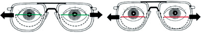
If, despite fully correcting the refractive error and achieving parallel alignment of the eyes for distance vision, a strabismic deviation persists at near and a high AC/A ratio is present, Franklin-type bifocal glasses should be used. They should only be used when there is potential for binocular vision; otherwise, their use is unjustified. As highlighted by Bredemeyer and Bullock [18], the purpose of bifocal spectacles is to provide a reduction in accommodative effort at near and to promote parallel alignment of the eyes. For this type of correction to function effectively, it is essential to position the segment line accurately in the center of the pupil. Correct and incorrect addition positioning in bifocal glasses is shown in Figure 1.
As Ansons and Davis [3] assert, the near deviation in strabismus might be more closely linked to proximal and tonic convergence than to accommodation. If strabismus is present at distance despite full correction, bifocal glasses should not be recommended [19]. Von Noorden and Campos [20] postulate similarly. Bifocal spectacles and miotic drugs are ineffective if the strabismic deviation at distance has not been completely eliminated. In PAET, prisms are used to restore binocular vision in situations where bifocal correction is insufficient to maintain binocularity, or as an alternative to surgery when surgical treatment is not feasible. In amblyopia management, occlusion therapy (patching) of the better-seeing eye is essential to improve visual acuity. Patching may influence the magnitude of strabismic deviation, particularly in non-accommodative esotropia.
Studies by Chun et al. [21] show that a significant reduction in the angle of strabismus is possible as a result of occlusion therapy (2-6 hours daily), ranging from 12 to 19 PD. Similarly, the studies by Koc et al. [22] in 63 participants demonstrated an improvement of an average of 11 PD over approximately one year of occlusion. Consequently, it is important to achieve the best possible visual acuity before making a decision about surgery, as the angle of strabismus may decrease as a result of improved visual acuity.
Vision therapy is possible to conduct in patients who have binocular vision that can be confirmed by subjective tests. Typically, patients with residual strabismus after surgery, who have the potential for binocular vision, are eligible for orthoptic therapy. Orthoptic exercises before the procedure require the responsibility of the orthoptist and the maturity of the patient, and should focus on the elimination of suppression and recognition of diplopia. The prerequisites for undertaking these exercises are the potential for binocular vision and sufficient control of eye alignment to achieve physiological diplopia at any distance; the possibility of improving negative vergence with the use of prisms [19] is also important. As emphasized by Molina-Martín et al. [23], passive therapy (spectacles and occlusion) combined with active therapy involving fusional vergence exercises is the most effective approach.
Botulinum toxin (BTA) serves as an adjunct treatment option, rarely used, which can help reduce small angles of strabismus and determine the fusion potential. It cannot replace surgery when indicated, as its effect is only temporary. In some centers, BTA is used prior to surgery in cases of mixed strabismus with a vertical component, in order to determine whether reducing the horizontal deviation will be sufficient to achieve binocular vision. The studies by Tejedor and Gutierrez-Carmona [24] suggest that BAT can be used before the target surgical procedure or, in some cases, such as PAET with a high AC/A, it may sometimes replace the surgical procedure or operation. As the authors emphasize, the key to effective botulinum toxin application is its injection at the appropriate time, typically around 5 years of age. In long-term use, it provides the greatest benefits in adults when binocular vision is present, and the angle of strabismus is unstable or surgery is undesirable [3].
Surgical treatment in PAET is indicated in cases of moderate to large angles of non-accommodative strabismus, where spectacle correction does not restore binocular function. The goal of the surgical procedure is to achieve alignment of the eyes in parallel in individuals with binocular potential or to maintain a residual angle of strabismus of 5-8 PD, i.e., approximately +4 degrees, in cases with a very low chance of binocular vision, in order to prevent, among other things, double vision.
Surgical treatment primarily reduces the non-accommodative component, which is why it is crucial to educate parents about the continued need for spectacle correction after strabismus surgery [20]. Beneficially, surgery may eliminate the need for bifocal spectacles, even in patients with a high AC/A ratio [14]. It is important to note that surgical intervention becomes particularly justified when concomitant oblique strabismus or alphabetic pattern is present. An associated vertical deviation exceeding 4 PD should be corrected to prevent adverse effects on binocular vision. There are various surgical techniques for the treatment of PAET. All techniques yield similarly satisfactory results when appropriately tailored to the patient’s needs and examination findings [25]. As highlighted by some authors, surgical intervention for late-onset accommodative esotropia w ith normal retinal correspondence may carry risks. Arnoldi [1] enumerates unfavorable factors associated with poorer postoperative outcomes in PAET. These include anisometropia greater than 0.75 DS, a spherical equivalent of more than +4.00 in the less hyperopic eye, weakened binocular vision, previous undercorrection of hyperopia, and a difference between distance and near deviation. In these cases, the risk of consecutive exotropia due to surgical overcorrection may be significantly elevated. In practice, many other factors are also considered, with the primary focus on achieving the greatest benefits for the patient. Studies by Kushner [26, 27] in 382 patients indicate that the overcorrection effect of surgery in individuals with a refractive error above +2.50 DS may be irreversible and may additionally result in pseudo-DHD (dissociated horizontal deviation). The most common complication following surgery for PAET is consecutive exotropia, while the rarest is cyclic esotropia [28]. As emphasized by Wright et al. [29], a 75% success rate of the procedure can be achieved if the prism adaptation test is performed beforehand. The test should be mandatory before surgical intervention in cases of PAET.
CONCLUSIONS
Convergent strabismus cases may present with ambiguous characteristics. In cases of partially accommodative esotropia, the non-accommodative component of esotropia persists despite full correction. As established by von Noorden and Campos [20], most patients, particularly those with infantile-onset esotropia, present with mixed-type strabismus − a finding corroborated by the author’s own clinical experience. The etiology of esotropia is primarily linked to uncorrected hyperopia and an elevated AC/A ratio. In the diagnostic workup for PAET, attention should be paid to the patient’s history, refraction testing after cycloplegia, assessment of visual acuity, ocular motility, cover test, accommodation and convergence status, AC/A ratio, and examination of the angle of strabismus and binocular functions. Among conservative treatment options, full refractive correction holds the most important role, followed by prismatic correction and vision therapy. Among the available invasive treatments, strabismus surgery and botulinum toxin application are the most frequently performed procedures. The highest potential for binocular vision in partially accommodative esotropia is noted in patients who developed strabismus after 1 year of age, received accurate diagnosis, and were treated promptly with appropriate therapy.


